

Hip tendon repair
Hip tendon repair primarily targets the tendons of the gluteus medius and minimus muscles, which attach to the greater trochanter on the side of the femur. When these tendons are torn or detached, they can disrupt pelvic stability and make certain movements difficult, such as walking or standing on one leg. The procedure typically involves reattaching the tendon to the bone using sutures and anchors.
Surgery is not always necessary. The decision takes into account the discomfort, the clinical examination, the extent and age of the lesion, the treatments already tried, and the patient's goals.
UNDERSTANDING TOTAL KNEE REPLACEMENT

-
This is the area located on the inner side of the knee and is the most frequently affected by osteoarthritis. Its wear and tear can cause internal pain and gradually lead to leg deformity.
-
Located on the outer side of the knee, this compartment is less frequently affected. Its wear and tear can cause lateral pain and gradually alter the leg's alignment.
-
This joint is located between the kneecap and the femur. When it is affected, pain often appears when climbing stairs, squatting, or after sitting for a long time.

WHEN TO CONSIDER A TOTAL KNEE REPLACEMENT?
Repair may be considered when a gluteal tendon injury causes significant and persistent discomfort, particularly if non-surgical treatments have not sufficiently improved function. Clinical examination and imaging must be interpreted together: a visible abnormality on an image is not, on its own, sufficient to decide on surgery.
- Complete rupture or avulsion of the tendon — Repair may be considered if the tendon is detached from its insertion and the injury corresponds to the symptoms and the examination.
- Marked weakness or functional impairment — Difficulty stabilizing the pelvis, walking, or performing certain activities may be a factor in the decision.
- Symptoms that persist despite treatment — Physiotherapy, activity adaptation and other situation-appropriate options are generally evaluated before considering surgery.
- Repairable damage and compatible lenses — The age of the rupture, its retraction, the quality of the tendon and the patient's expectations are taken into account to estimate the possible benefit.
Tendinopathy or a partial tear does not automatically lead to surgery. The decision is made on a case-by-case basis, after examining the expected benefits, the constraints of rehabilitation, and non-surgical solutions.
PREPARE FOR THE INTERVENTION
The preparation helps to confirm the diagnosis, plan the repair and organize the period during which weight-bearing and certain movements may be limited.
PRE-OPERATIVE ASSESSMENT
The surgeon will review your symptoms and medical history, examine your hip, and review any available imaging studies. Further assessments and an anesthesia consultation will be scheduled based on your health status and the hospital's practices.
INTERVENTION PLANNING
The technique depends on the location and extent of the tear, tendon retraction, and tissue quality. The surgeon determines the type of reinsertion and the number of anchors based on the findings and the necessary procedure.
PHYSICAL PREPARATION AND PRACTICE
Pre-operative exercises are tailored to the pain and the injury. It is helpful to plan for your return home, clear walkways, and arrange for temporary assistance if mobility is difficult. Crutches may be necessary after the operation.
INSTRUCTIONS BEFORE THE OPERATION
The team will provide you with instructions regarding fasting, hygiene, and arrival at the clinic. Do not change or stop any medication without consulting the medical team or the anesthesiologist.

HOW DOES THE PROCEDURE WORK?
The procedure depends on the rupture and the technique used. Repair can be performed endoscopically, through small incisions, or via an open approach. The goal is to reposition the tendon over its insertion point and fix it securely.
INSTALLATION AND ANESTHESIA
The procedure is performed in the operating room, under general anesthesia or another anesthetic strategy decided upon with the anesthesia team. After the patient is positioned, the skin is prepared, and the surgical field is disinfected and covered with sterile drapes. The team checks, in particular, the side to be operated on and the patient's file before beginning.
Access to the treated area
For endoscopic repair, the surgeon makes several small incisions to insert a camera and instruments around the greater trochanter. For open repair, a larger incision provides direct access to the tendons. The surgical approach depends on the injury and the necessary exposure.
Main surgical procedure
The surgeon examines the rupture, identifies the affected tissues, and releases the tendon if necessary so that it can return to its insertion without excessive tension. The bone attachment area may be prepared to expose a surface conducive to healing. Anchors are then placed in the greater trochanter. The sutures attached to these anchors are passed through the tendon and then knotted or fixed according to the technique used to bring the tendon closer to the bone. In cases of extensive injury or insufficient tissue quality, reinforcement or an additional procedure may be considered; it is not necessary in all repairs.
result check
The surgeon checks that the tendon is reattached to its insertion, that the fixation is secure, and that the repair remains consistent during controlled hip movements. He also takes care to preserve the surrounding structures.
Closure and end of the intervention
The incisions are closed and covered with a dressing. A splint may be prescribed in some situations, but it is not always necessary. The patient is then taken to the recovery room for monitoring after the anesthesia wears off.
The repair must then be protected during the healing period: weight-bearing and movements allowed follow the surgeon's personalized instructions.
The duration of the operation and the type of hospitalization vary depending on the extent of the injury, the surgical technique, and the patient's medical condition. Some teams report procedures lasting one to three hours and a possible overnight hospital stay, but these guidelines do not predetermine the specific arrangements made for each patient.
THE DIFFERENT POSSIBLE TECHNIQUES
The surgical approach is chosen according to the size and location of the tear, its retraction, and the quality of the tendons. Endoscopic and open repairs most often use sutures and anchors to reattach the tendon to the bone.
The choice of technique aims to achieve a repair tailored to the injury while allowing for protective rehabilitation. Additional options are considered only if the surgical findings warrant them.
Endoscopic repair
A camera and instruments are inserted through small incisions to repair the tendon. This approach limits the size of the incisions, but it is not suitable for all ruptures, particularly when direct access or a more extensive repair is required.
Repair via open access
An incision allows direct exposure of the tendon and its insertion. This approach may be chosen for certain large, retracted, or complex tears. It provides direct access, but with a larger incision and the usual limitations of open surgery.
Fixation using anchors and sutures
The anchors are implanted in the greater trochanter and have sutures that secure the tendon to the bone. Their number and placement depend on the size of the tear and the available tissue. The surgeon selects the appropriate material for each case.
Reinforcement or associated gesture
Reinforcement may sometimes be considered when the tendon is fragile or the tear extensive. Other procedures may be combined depending on the findings, but they are not always necessary. The benefit of reinforcement must be weighed against the added complexity of the procedure.
The method chosen depends on the anatomy and the lesion, and not on a single technique applicable to all.
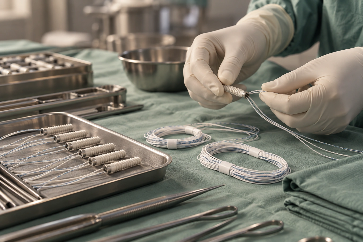
AFTER THE PROCEDURE: THE FIRST STEPS
Recovery room and pain
Monitoring focuses on waking up after anesthesia, comfort, and overall condition. Pain medication is provided as prescribed by the medical team. Pain may change in the first few days; report any poorly controlled pain to the healthcare staff.
Support, walking and splint
Weight-bearing may be limited initially to protect the repair. Crutches are often necessary; a splint may be prescribed depending on the procedure. The amount of weight-bearing allowed and movements to avoid are determined by the surgeon and physiotherapist. Protocols vary depending on the repair and the medical team: some involve limiting weight-bearing for several weeks.
Return home and monitoring
The procedure may be performed on an outpatient basis or include an overnight stay, depending on the procedure, the patient's health, and their ability to move around safely. Keep the dressing clean and follow the aftercare instructions provided. Contact the team if you develop a fever, significant drainage, spreading redness, a marked increase in pain, or any unusual symptoms. The first follow-up appointment will allow us to assess the patient's progress and determine their recovery.
REHABILITATION AND RETURN TO ACTIVITIES
In the first few weeks, the priority is to protect the fixation and adhere to the weight-bearing and movement guidelines. Depending on the repair, certain movements—particularly active leg abduction—may be temporarily limited. The schedule is determined by the surgeon and physiotherapist.
Rehabilitation then progresses in stages: controlled recovery of mobility, muscle activation, strengthening of the hip and trunk muscles, balance, and pelvic control. The exercises are adapted to the healing process and the hip's response.
Walking, climbing stairs, driving, and returning to work depend on weight-bearing restrictions, the use of crutches, mobility, and the demands of the job. A sedentary job and a physically demanding job do not have the same constraints. Driving can be resumed when aids are no longer needed, reflexes are sufficient, and the medical team gives the go-ahead.
A return to sports is gradual, following medical clearance, taking into account strength, mobility, and movement control. Timeframes vary depending on the repair and progress: some hospital documents mention several months before a return to demanding sports, but this is not a guaranteed timeline.
The repair heals gradually: respecting the rehabilitation stages is part of the treatment.

EXPECTED RESULTS, LIMITATIONS AND RISKS
OBJECTIVES AND EXPECTED RESULTS
The procedure aims to reattach the tendon to the bone, reduce discomfort caused by the rupture, and improve hip strength and stability. Results depend on factors such as the extent of the injury, the condition of the tendon, and the healing process.
BOUNDARIES
Repair does not guarantee the disappearance of all pain or a return to previous function. Weakness, stiffness, or discomfort may persist, particularly if the rupture is old or if the tendon is severely retracted or damaged.
RISKS AND COMPLICATIONS
Like any surgical procedure, it carries risks, including infection, hematoma, phlebitis, wound healing problems, or persistent pain. The repair may not heal sufficiently, may detach, or may rupture again; further surgery may then be considered. Neurological or vascular complications are possible but rare.
The expected benefit must always be assessed in light of the limitations of the repair, the operative risks and the constraints of rehabilitation. The risks of non-healing and further surgery are described in particular in hospital information intended for patients.

AN INTERVENTION TAILORED TO EACH PATIENT
Two patients with a lesion that appears similar on imaging may have different symptoms, strength, and needs. Therefore, they do not necessarily require the same strategy.
THE Dr. Benoît Coulin It takes into account age, anatomy, tissue quality, the severity and age of the injury, activity level, occupation, associated injuries, and treatments already tried. Personal goals—walking more comfortably, resuming activity, or regaining better stability—also play a role in the decision.
The surgical approach, the repair procedure, the protective measures, and the recovery program are defined based on these factors. The discussion also allows for a comparison of the expected benefits of surgery with the continuation of non-surgical treatment.
"To move is to live, it is freedom. We must do everything to give life back, one patient after another."
FREQUENTLY ASKED QUESTIONS
Is hip tendon repair appropriate for your situation?
A clinical examination and imaging analysis help to define the lesion, evaluate the options and discuss the technique best suited to your objectives.
