

FEMORAPATELLARY SYNDROME
Patellofemoral pain syndrome refers to pain located at the front of the knee, around or behind the kneecap. It often occurs during activities that put stress on the bent knee, such as going up or down stairs, running, or squatting.
This pain does not necessarily mean that the cartilage is damaged or that the kneecap is displaced. A clinical examination can determine the cause and allow for appropriate treatment, most often without surgery.
UNDERSTANDING PATELLAR FEMOROSYNDROME

-
This is the area located on the inner side of the knee and is the most frequently affected by osteoarthritis. Its wear and tear can cause internal pain and gradually lead to leg deformity.
-
Located on the outer side of the knee, this compartment is less frequently affected. Its wear and tear can cause lateral pain and gradually alter the leg's alignment.
-
This joint is located between the kneecap and the femur. When it is affected, pain often appears when climbing stairs, squatting, or after sitting for a long time.
RECOGNIZING THE SYMPTOMS
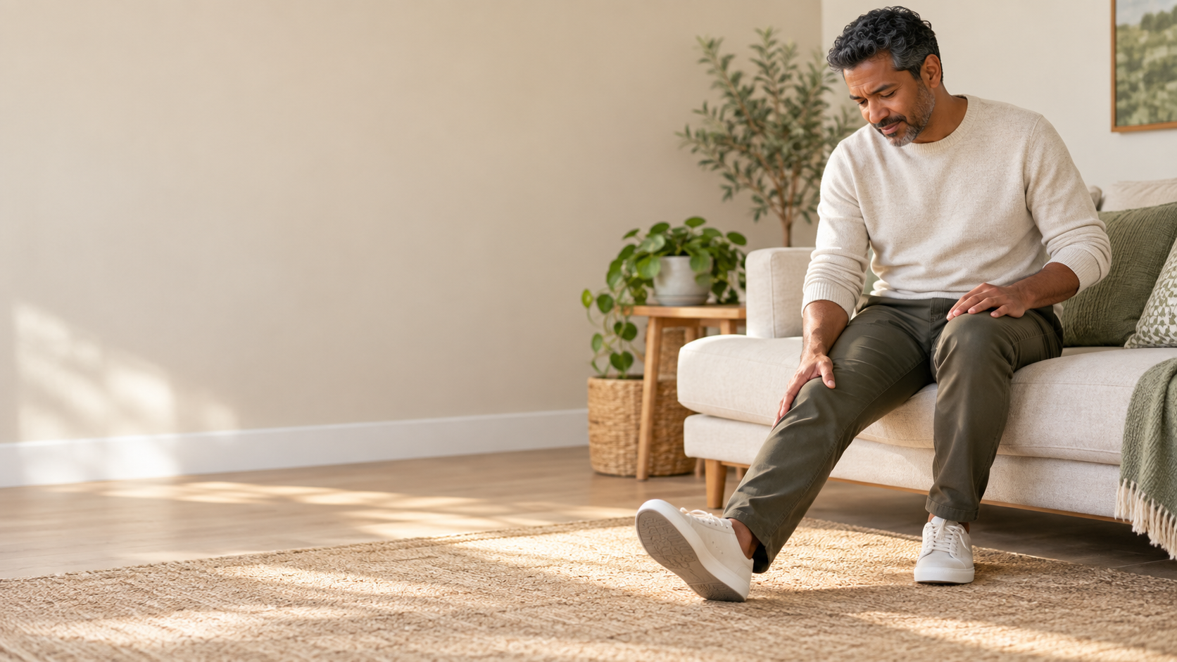
The pain is usually felt at the front of the knee, around or behind the kneecap. It can vary depending on the activities and their intensity.
- Pain on the stairs — The discomfort is often more noticeable on the descent, when the knee is bent and the muscles are slowing the movement.
- Pain when sitting for extended periods — A knee that is kept bent for a certain time can become painful or stiff, with discomfort when standing up.
- Pain when running or jumping — Activities with repeated impacts or changes in pace can trigger symptoms.
- Discomfort when squatting — Bending the knee sharply or rising from a low position can increase the pressure exerted on the patellofemoral joint.
- Cracking or rubbing sensations — They can accompany the movements, but do not by themselves mean that there is a significant injury.
Pain can develop gradually after increasing training, resuming sports, or changing the type of activity. If it persists, recurs regularly, or limits daily movements, an evaluation can help identify the cause. In cases of pain after an impact, rapid swelling, inability to bear weight, or a locked knee, seek prompt evaluation from [medical professional/organization name]. SOS Knee Geneva.
Why does patellofemoral pain syndrome occur?
Patellofemoral pain syndrome is characterized by pain in the area between the kneecap and the femur. Its onset is often linked to several factors that alter the load borne by the knee. Pain alone is not sufficient to diagnose an abnormality of the kneecap or cartilage.
Too rapid an increase in demand
A sudden increase in the volume or intensity of running, jumping, or training can exceed the knee's ability to adapt. Changes in terrain, footwear, or training frequency can also contribute to the onset of symptoms.
Movement control
The way the knee bends and straightens, as well as hip and pelvic control during weight-bearing, can influence the stresses around the kneecap. Weak or insufficient muscle endurance can make certain movements more difficult to control, but it is not necessarily the sole cause of pain.
Anatomical features and history
The shape of the patella or trochlea, its height, the alignment of the lower limb, or a previous dislocation can sometimes contribute to the symptoms. These factors must be interpreted in conjunction with the clinical examination: a visible anatomical peculiarity does not always explain the pain.
Patellofemoral syndrome should be distinguished from’patellofemoral osteoarthritis, This corresponds to damage to the cartilage and the joint between the kneecap and the femur. Both conditions can cause pain at the front of the knee, but their diagnosis and treatment are not the same.
MAKE THE RIGHT DIAGNOSIS
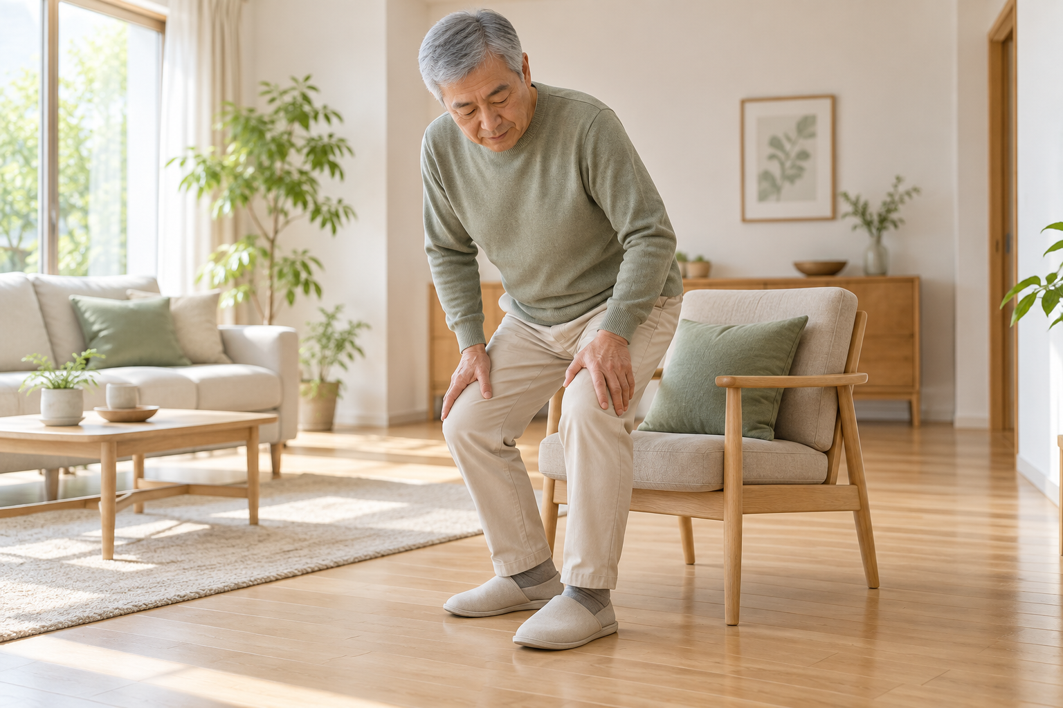
The diagnosis is initially based on symptoms and examination of the knee. Imaging is chosen according to the context and the observed findings.
01 — Circumstances and evolution
Dr. Benoît Coulin specifies the location of the pain, the activities that trigger it, its duration, and any recent changes in training or activity. A history of trauma or dislocation is also sought.
02 — Clinical examination
The examination assesses knee mobility, the patella, tender areas, strength, and lower limb control during functional movements. It also helps to identify other possible causes of pain.
03 — X-rays if they are useful
X-rays may be recommended if the examination, medical history, or persistent symptoms warrant it. They help assess the alignment and shape of bone structures, as well as detect certain joint conditions.
04 — MRI or other targeted examination
An MRI is not always necessary. It may be requested when symptoms persist, after trauma, or if the examination suggests cartilage damage or another condition requiring further investigation. A CT scan can sometimes be useful to analyze certain bone structures or patellar alignment.
Pain guides the assessment; imaging is never interpreted alone.
Initial care
Initially, the goal is to reduce the stresses that clearly aggravate the pain, while maintaining movements and activities that remain comfortable.
Temporarily adapt activities
It may be helpful to temporarily reduce running, jumping, repetitive descents, or deep bending if these activities worsen symptoms. Adaptation depends on the knee's response: it is not necessary to stop all activity if some movements remain well tolerated.
Gradually resume
A gradual return to activity helps avoid increasing the workload too quickly. The duration, frequency, terrain, and intensity can be adjusted according to the evolution of pain during the activity and in the hours that follow.
Relieving the symptoms
Applying cold compresses can provide temporary relief if the knee is sore after exercise. Pain relievers or anti-inflammatory medications are not suitable for everyone and should be considered based on medical history and the advice of a healthcare professional.
A prompt consultation is recommended if the pain follows a significant trauma or is accompanied by marked swelling, locking, or difficulty bearing weight. SOS Knee Geneva offers an assessment of knee problems requiring prompt attention.

Conservative treatment
Treatment for patellofemoral pain syndrome is generally conservative. Surgery is not routine and is not usually indicated for isolated patellofemoral pain without structural abnormalities or specific instability.
Physiotherapy and exercise progression
Physiotherapy can help gradually improve the strength and endurance of the thigh and hip muscles, as well as movement control. The program is tailored to the patient's abilities and the movements that trigger their symptoms.
Load adjustment
Activity is reintroduced gradually, taking into account the knee's reaction. The physiotherapist can help organize a gradual return to brisk walking, running, or exercises that involve the knee being bent.
Monitoring and reassessment
Follow-up allows for monitoring the evolution of symptoms and adjusting exercises or activities. A reassessment is useful if the pain does not improve despite consistent treatment, if it worsens, or if new symptoms appear. Further evaluation can then be conducted to rule out other underlying causes.
Progress is determined by the function and reaction of the knee, not by an imposed schedule.
To better understand the possible options in case of persistent patellar problems, see the page on the Patellar surgery and its indications.
surgical treatment
Surgery is rarely indicated for isolated patellofemoral pain syndrome. It may be considered if an assessment reveals a specific anatomical cause, recurrent patellar instability, or an associated injury, and if symptoms persist despite appropriate conservative management.
Depending on the identified cause, options may include patellar stabilization, patellar path correction, or a procedure targeting an associated lesion. The choice depends on the symptoms, examination, and imaging results; no single technique is suitable for all patients.
The decision to intervene is made after confirming the diagnosis and assessing the response to conservative treatment. When surgery is considered, Dr. Benoît Coulin explains the expected outcomes, the limitations of the procedure, and the rehabilitation steps. The goal is to improve function or stability when indicated, without promising the complete elimination of all pain.
For more details, see the indications and techniques of patellar surgery.

Rehabilitation and resumption of activities
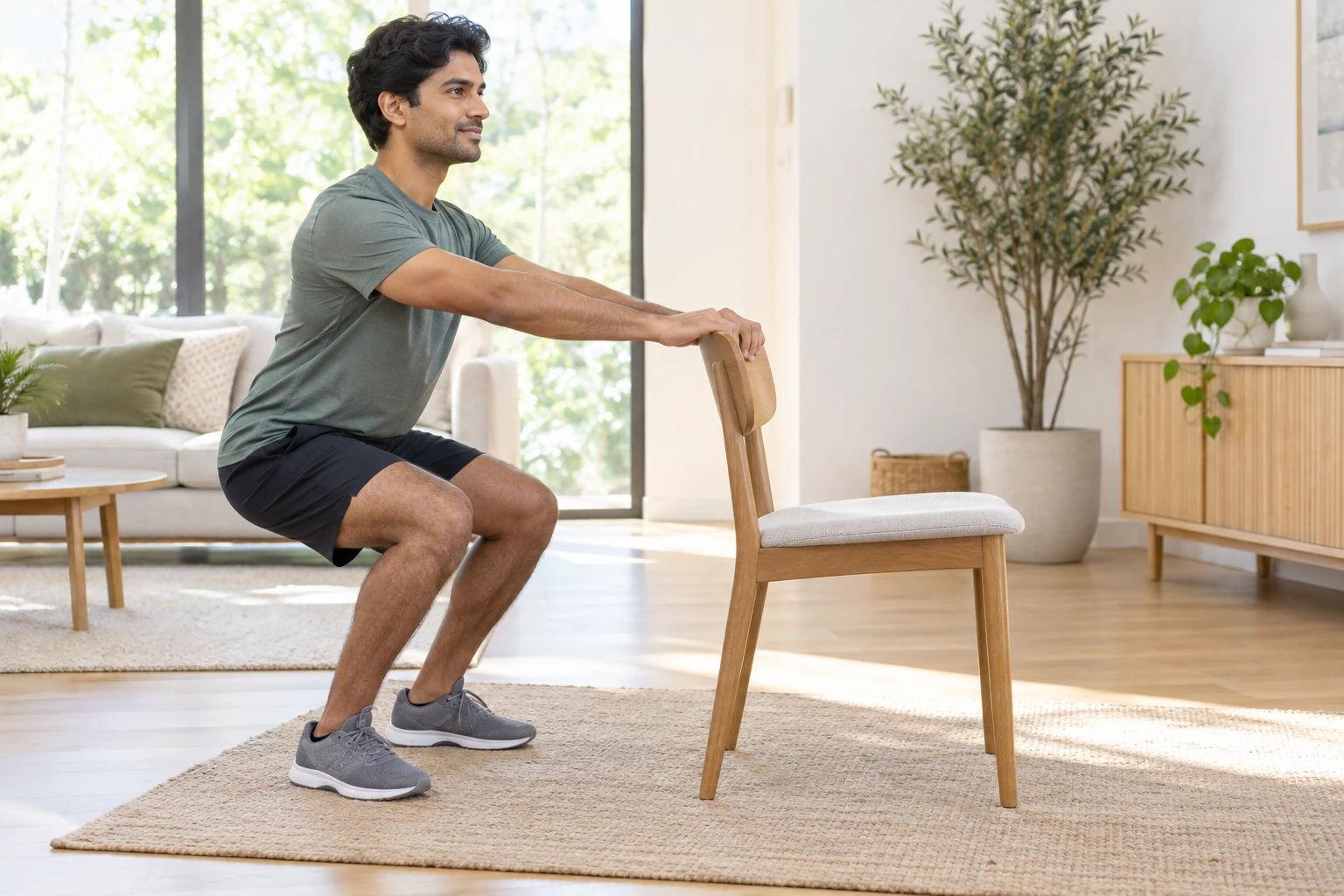
Recovery is gradual, depending on pain, mobility, strength, and knee control. The steps and timelines vary depending on the individual and any associated injuries.
01 — Maintain comfortable mobility
Knee movements are maintained within a tolerated range of motion. Exercises are adjusted if significant flexion causes marked pain.
02 — Rebuilding strength
Strengthening of the thigh and hip muscles progresses according to tolerance. The load increases when exercises are performed with satisfactory control and without sustained worsening of symptoms.
03 — Improve movement control
Functional work can focus on balance, support and knee control during movements such as sitting down, standing up or climbing a step.
04 — Resume daily activities
Walking and usual activities are increased according to comfort and knee response. Swelling or a persistent increase in pain may lead to a temporary reduction in load.
05 — Gradually resume sports
Running and activities involving jumping or changes of direction are reintroduced when strength, control, and tolerance to exertion are sufficient. Functional tests and clinical progress can guide this step.
There knee rehabilitation after patellar surgery It is planned according to each patient's situation.
The recovery is guided by the regained abilities and the knee's response to exertion.
Care tailored to each patient
Two people experiencing pain in the front of the knee may have different symptoms and causes. Dr. Benoît Coulin considers the location of the pain, its progression, the activities involved, the patient's medical history, the clinical examination, and the results of any necessary tests.
Physiotherapy and activity adaptation are often preferred, but their content must be tailored to the patient. They require steady progress and may take time. Intervention is only considered if the assessment reveals a specific indication and conservative options do not adequately address the identified problem.
«"Understanding the cause of the pain allows us to choose a progression adapted to each individual's knee and activities."»
FREQUENTLY ASKED QUESTIONS
Does your pain in the front of your knee persist?
A clinical examination and, if necessary, appropriate imaging can help to distinguish patellofemoral pain syndrome from other causes of pain and to define appropriate management.
