

Focal lesion of the cartilage
A focal cartilage lesion corresponds to a well-defined area where the joint lining is damaged. It can cause pain or swelling, while the surrounding cartilage remains intact.
The goal is to understand the origin of the symptoms and to choose the solutions that will allow relieve knee pain while preserving its function.
The goal of care is first to understand the origin of the symptoms, then to choose solutions to preserve movement and autonomy.
UNDERSTANDING THE FOCAL LESION OF THE CARTILAGE

-
This smooth tissue covers the ends of the bones. It facilitates the gliding of surfaces and helps to distribute pressure within the joint.
-
This rounded part of the femur articulates with the tibia. Its cartilaginous surface may have a localized defect in an area subjected to weight-bearing.
-
The cartilage is cracked, detached, or absent in a specific area. The lesion may be superficial or extend through its entire thickness.
-
Located beneath the cartilage, it provides support for it. If this bone is also affected, it is called an osteochondral lesion.

RECOGNIZING THE SYMPTOMS
A focal cartilage lesion can become painful after trauma or develop gradually. The discomfort varies depending on its location, depth, and any associated injuries.
- Pain during exertion: Prolonged walking, running, stairs or certain bending movements can trigger pain.
- Swelling : The knee may swell after activity or experience repeated episodes of effusion.
- Hookups: An irregular surface or a loose fragment can impede movement, sometimes to the point of blocking.
- Loss of mobility: Fully bending or straightening the knee can become uncomfortable, especially when it is swollen.
These symptoms can also originate from the meniscus, ligaments, or kneecap. An assessment helps to identify its true origin.
WHY DOES CARTILAGE DETERIORATE?
A focal cartilage lesion can result from an accident or repeated mechanical stress. The assessment aims to determine the cause of the damage and any factors that may contribute to its perpetuation.
TRAUMA
A twist, impact, or dislocation of the kneecap can damage the cartilage. This injury may accompany a ligament or meniscal tear.
LEG AXIS
Misalignment can concentrate pressure on one part of the knee and promote localized overload of the joint surface.
INSTABILITY
Abnormal movements in an unstable knee can increase stress on the cartilage. Therefore, the ligaments and the patellar guidance system are examined.
BONE DAMAGE
Certain conditions affecting the subchondral bone, such as osteochondritis dissecans, can weaken the cartilage above and create an osteochondral lesion.
MAKE THE RIGHT DIAGNOSIS
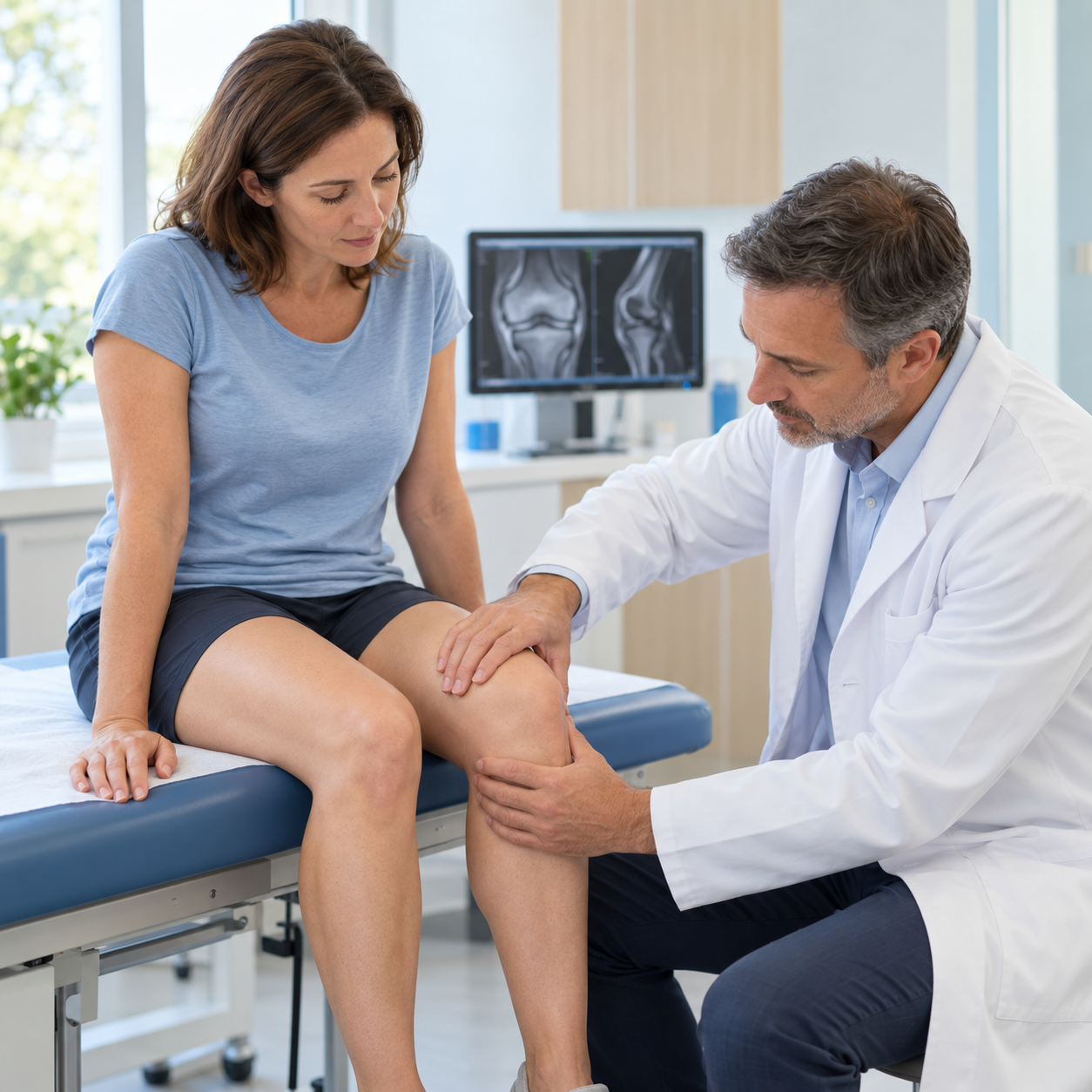
The assessment of a focal cartilage lesion combines symptoms, examination of the knee, and appropriate imaging. It specifies the characteristics of the defect and its impact on activities.
01 — THE CLINICAL EXAMINATION
Pain, swelling, mobility, stability, and leg alignment are analyzed. The circumstances of onset and the patient's goals also guide the assessment.
02 — X-RAYS IN CHARGE
They assess alignment, joint space, and bone changes. A normal X-ray does not rule out an injury, as cartilage is not directly visible on it.
03 — ADDITIONAL EXAMINATIONS
MRI helps to determine the extent, depth, and location of the lesion. It also looks for damage to the underlying bone, menisci, or ligaments.
A lesion visible on an MRI is not enough to decide on surgery. The discomfort experienced and the functioning of the knee remain crucial factors.
RELIEVING KNEE PAIN WITHOUT SURGERY
When there is no immediate indication for surgery, treatment of a focal cartilage lesion can begin with conservative management.
The goal is to reduce pain, improve support, and maintain activity that is compatible with the condition of the knee.
PHYSICAL ACTIVITY AND PHYSIOTHERAPY
Physiotherapy focuses on strengthening the thigh and hip, improving knee mobility and control. Exercises are tailored to the location of the injury and the joint's response.
ADAPTATION OF DAILY LIFE
Temporarily reducing impact or painful movements can improve comfort. Alternating between activity and recovery allows for better management of stress.
When excess weight is present, addressing it can also help to reduce the load borne by the knee.
DRUG TREATMENTS
Pain relievers or, in certain situations, anti-inflammatories may be offered depending on the patient's history and contraindications.
They act on the symptoms, without rebuilding cartilage or replacing rehabilitation.
KNEE INFILTRATIONS
An injection may sometimes be considered to improve symptoms. Its usefulness depends on the context and the product used: not all injections are suitable for an isolated focal lesion.
A decrease in pain does not mean that the cartilage defect is repaired.
Preserving movement and improving comfort remain the primary objectives when non-surgical treatment is possible.

WHEN TO CONSIDER SURGERY?
Surgery may be considered when pain and limitations persist despite appropriate treatments. An unstable fragment or mechanical blockage may also warrant surgical intervention.
The choice depends on the size and depth of the lesion, the condition of the underlying bone, the stability of the knee, and the patient's activities.
REGULARIZING: CHONDROPLASTY
Chondroplasty allows for the smoothing of unstable edges or the removal of a troublesome fragment, often arthroscopically. It aims to improve mechanical symptoms without recreating the original cartilage.
STIMULATE: MICROFRACTURES
Small perforations in the bone beneath the lesion promote the formation of repair tissue. This tissue, being less resistant than the original cartilage, limits its use to certain lesions and patient profiles.
RESTORATION: GRAFTS AND MATRIXES
Osteochondral grafting, cell therapy, or matrix augmentation may be considered to restore a deficient area. These procedures require precise selection, with possible correction of axial deformities or instability.
KEEP MOVING WITH A CARTILAGE INJURY
A focal cartilage lesion does not necessarily require the cessation of all activity. Appropriate movement helps maintain muscle strength and mobility.
Low-resistance cycling, swimming, or strengthening exercises can be recommended when well tolerated. The choice depends on the symptoms and the location of the injury.
Increasing pain, repeated swelling, or loss of mobility after exertion should lead to adjustments in activity and a reassessment of the situation.
After reconstructive surgery, weight-bearing and certain movements may be temporarily limited. Returning to sports follows a gradual program and may take several months.
The activity must follow the capabilities of the knee and the stages of treatment.


A CARE APPROACH TAILORED TO EACH PATIENT
There is no single treatment for focal cartilage lesions. Two defects of similar size can have different consequences depending on their location and the patient's needs.
THE Dr. Benoît Coulin A gradual approach based on a thorough assessment is preferred. Conservative treatments are considered when appropriate to the situation.
If an operation is necessary, its objective is defined according to the problem encountered: to treat a troublesome fragment, to promote a repair or to restore a deficient area.
Age, professional activity and sporting projects all play a role in the decision.
«"Treating an area of damaged cartilage also helps preserve the balance and movement of the entire knee."»