

Osteochondral grafting and mosaicplasty
Osteochondral grafting involves transferring one or more cylinders of cartilage and bone to a damaged joint area. When several small grafts are placed side by side, the technique is called mosaicplasty. It can be considered for certain focal lesions in a joint whose overall condition allows for this procedure.
The indication for surgery depends on the functional impairment, the clinical examination, the observed lesions, the treatments already tried, and the patient's goals. An abnormal image alone is not sufficient to decide on surgery.
UNDERSTANDING PARTIAL KNEE REPLACEMENT

-
This is the area located on the inner side of the knee and is the most frequently affected by osteoarthritis. Its wear and tear can cause internal pain and gradually lead to leg deformity.
-
Located on the outer side of the knee, this compartment is less frequently affected. Its wear and tear can cause lateral pain and gradually alter the leg's alignment.
-
This joint is located between the kneecap and the femur. When it is affected, pain often appears when climbing stairs, squatting, or after sitting for a long time.

WHEN TO CONSIDER AN OSTEOCHONDRAL TRANSPLANT?
Osteochondral grafting may be considered when a localized lesion of the cartilage and underlying bone causes persistent discomfort, and when assessment indicates that this technique is suitable. The indications are evaluated on a case-by-case basis, as the harvesting area is limited by the available tissue within the knee.
- Symptomatic focal lesion: The discomfort remains present and localized despite appropriate treatment.
- Osteochondral involvement: The assessment shows that the cartilage and possibly the underlying bone are involved.
- Persistent functional limitation: The symptoms interfere with daily, professional, or sporting activities.
- Results compatible with the technique: The location and extent of the lesion allow for the consideration of one or more grafts.
The decision also takes into account functional age, activity level, associated injuries, and the patient's goals. Transplantation is not automatically necessary: non-surgical treatment or another option may be preferable depending on the assessment.
To learn more about knee cartilage damage, consult the page dedicated to knee cartilage lesions on SOS Genou Genève.
PREPARE FOR THE INTERVENTION
The preparation phase allows for confirmation of the surgical plan, discussion of the anesthesia, and organization of the initial post-operative steps. Examinations and instructions are tailored to each individual situation.
PRE-OPERATIVE ASSESSMENT
The surgeon reviews the symptoms, examines the knee, and reviews any previously performed tests. Further tests or analyses may be ordered. The anesthesia consultation assesses the patient's medical history and anesthesia options.
INTERVENTION PLANNING
The size and location of the lesion help determine whether one or more grafts can be used and which surgical approach is appropriate. The plan can be adjusted based on findings during the procedure.
PHYSICAL PREPARATION AND PRACTICE
The team may advise maintaining appropriate activity levels before the operation. At home, it is helpful to plan your movements in advance and arrange for assistance if weight-bearing instructions temporarily limit walking. Crutches will be prepared if prescribed.
INSTRUCTIONS BEFORE THE OPERATION
The team will specify the fasting requirements, hygiene guidelines, and arrival time. Inform the team of all your medications and do not discontinue them without consulting the medical team.
For practical information on organizing knee surgery, consult the "Preparing for your operation" page of SOS Knee Geneva.
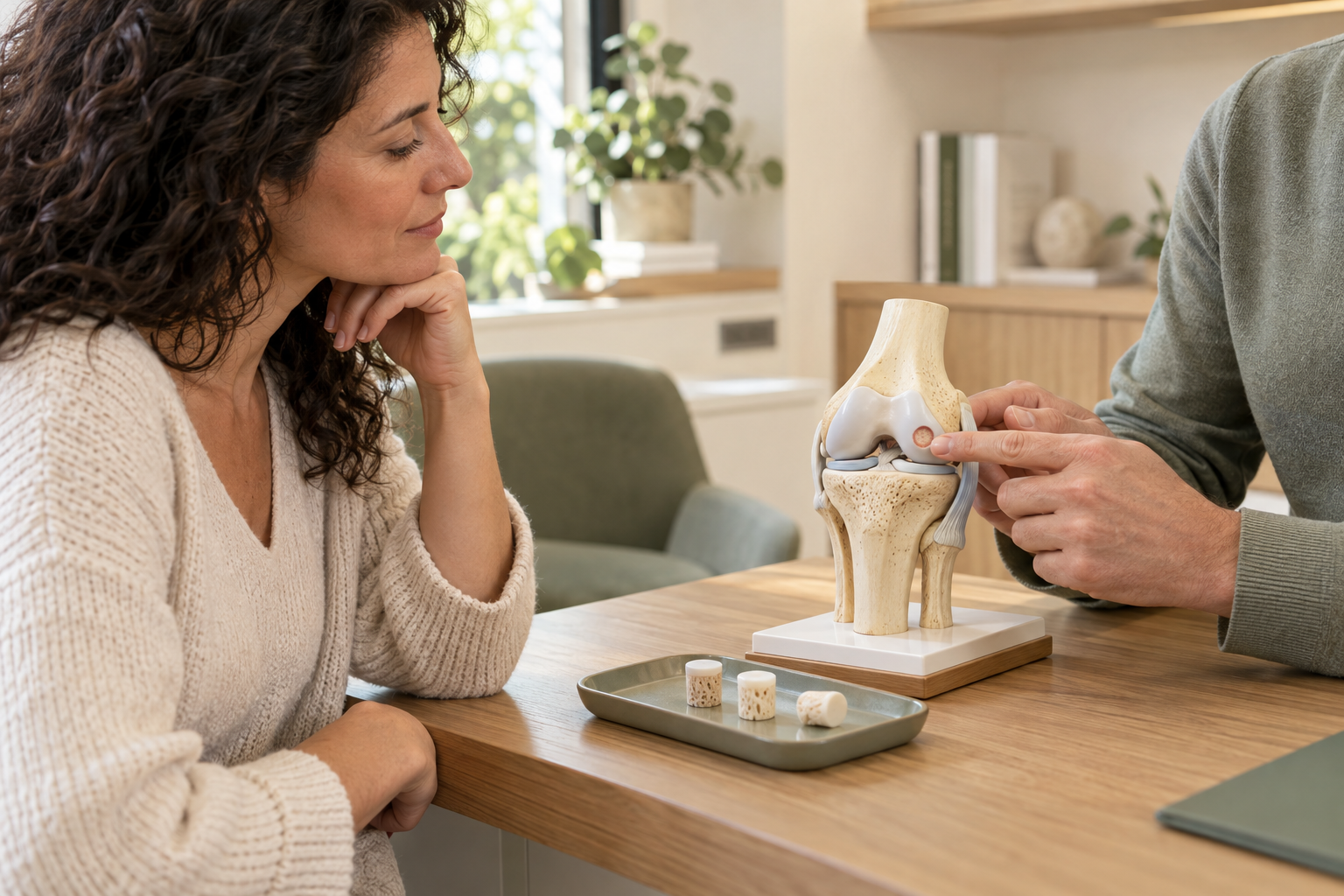
HOW IS AN OSTEOCHONDRAL TRANSPLANT PERFORMED?
The procedure involves preparing the affected area, harvesting the graft(s), and then positioning them in the targeted area. The approach and exact steps depend on the location and size of the lesion.
INSTALLATION AND ANESTHESIA
The patient is positioned on the operating table. General or regional anesthesia may be considered depending on the medical situation and the consultation with the anesthesiologist. The team prepares the skin under sterile conditions and verifies the patient's identity, the side to be operated on, and the planned procedure before the start of the operation.
Access to the treated area
Depending on the injury and the chosen technique, the procedure may require arthroscopy, targeted opening surgery, or a combination of both. The arthroscope allows visualization of the inside of the joint through small incisions. The chosen approach must allow for precise work on the area to be treated and the donor site.
MAIN SURGICAL PROCEDURE
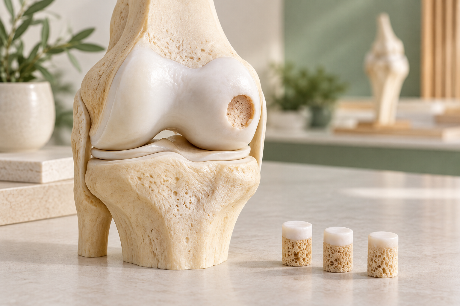
The surgeon assesses the injury and prepares the recipient site. One or more cylinders of cartilage and bone are harvested from a less stressed area of the same knee. Each graft is then trimmed and placed in the damaged area. For mosaicplasty, several small grafts can be positioned side by side to cover the target area. This technique depends on the amount of available tissue and the shape of the injury; therefore, it is not suitable for all conditions.
RESULT CONTROL
The surgeon checks the position and stability of the grafts as well as the regularity of the joint surface. He also monitors the mobility accessible during the procedure and takes care to preserve the surrounding structures.
CLOSURE AND END OF THE PROCEDURE
The instruments are removed, the incisions are closed, and a dressing is applied. A splint may be used if the chosen protocol requires it. The patient is then transferred to the recovery room for monitoring.
The goal is to position the graft(s) stably while respecting the knee tissues.
The duration varies depending on the lesion, the number of grafts, and the surgical approach. Outpatient treatment may be possible in some cases, while hospitalization may be necessary depending on the procedure and the patient's condition. The team will confirm these arrangements before the operation.
WHAT TECHNIQUE IS USED FOR OSTEOCHONDRAL TRANSPLANTATION?
Mosaicplasty is a form of osteochondral autograft: the tissue used comes from the same knee. A single graft or several cylinders can be considered depending on the shape and extent of the lesion.
The choice does not depend solely on the size of the area. The location, the condition of the surrounding cartilage, the underlying bone, and the patient's goals all contribute to the decision. The AAOS describes the transfer of a single graft or multiple grafts, known as mosaicplasty, and emphasizes that autografting is limited by the amount of available tissue.
Single graft
A single osteochondral cylinder can be used when it matches the shape of the lesion. This option avoids multiplying the sampling sites, but does not allow for coverage of all surfaces.
Mosaicplasty
Several small grafts are placed side by side to cover a targeted area. This technique can be adapted to certain types of lesions, but it remains limited by the quantity of graft available and by the constraints of the donor site.
Arthroscopy or targeted approach
Arthroscopy uses a camera and instruments inserted through small incisions. Depending on the location of the lesion, a targeted opening can facilitate access and graft placement. The choice depends on the required visibility and access.
Graft stabilization
The grafts are adjusted and positioned in the prepared area. Stability is checked during the procedure. The precise methods depend on the graft and the findings during the operation; no fixation device is systematically used.
The chosen technique is the one that best suits the injury, the anatomy of the knee and the patient's goals.
AFTER THE PROCEDURE: THE FIRST STEPS
In the recovery room, the team monitors the patient's overall condition, knee, and pain. Initial checks and pain management are tailored to the procedure and the individual patient.
Instructions regarding support and mobilization protect the graft(s) during the initial stages of recovery. These instructions are provided before discharge and may change during follow-up.
WAKE-UP AND PAIN ROOM
The team monitors vital signs, pain levels, and the dressing. Discomfort or swelling may occur. Medications and comfort measures are prescribed as needed.
SUPPORT, WALKING AND SPLINTING
Weight-bearing can be protected, limited, or gradual depending on the technique. Crutches are used if prescribed. A splint is not always necessary; it depends on the surgical instructions. Initial movements are performed within the limits set by the surgeon.
RETURN HOME AND MONITORING
Discharge may occur the same day or after a short hospital stay, depending on the procedure and the patient's condition. The team will provide instructions for dressing changes and the initial check-up. Contact the team immediately if you experience fever, significantly worsening pain, redness or drainage from the incision, or a painful and swollen calf. These signs are among those that warrant medical attention after knee surgery.
REHABILITATION AFTER AN OSTEOCHONDRALE TRANSPLANT
Initially, rehabilitation follows guidelines for graft protection. The primary objectives are pain and swelling control, wound healing, permitted mobility, and muscle reactivation. Weight-bearing remains within the surgeon's instructions.
The progression can then work on mobility, strength, balance, and movement control. The exercises are adapted to the knee's development and the patient's tolerance.
Walking, climbing stairs, driving, and work resume according to functional abilities, support guidelines, and the type of professional activity. Sedentary work and physical work do not impose the same constraints.
Sports activities are resumed gradually, after medical clearance and depending on healing, mobility, strength, and knee control. Timeframes vary; no schedule can be guaranteed in advance.
The resumption of activities depends on the progress of the knee and adherence to graft protection guidelines.
To discover the areas of knee care, consult the SOS Knee Geneva website.

EXPECTED RESULTS, LIMITATIONS AND RISKS
Objectives and expected results
The treatment aims to restore a localized area of the joint surface and reduce the discomfort associated with the lesion. Functional improvement is sought, but a complete result or the absence of pain cannot be guaranteed.
Boundaries
Recovery varies depending on the extent and location of the injury, the overall condition of the knee, and the progress of rehabilitation. Discomfort may persist at the donor site. The amount of available tissue limits the indications for osteochondral autografting.
Risks and complications
Like any surgical procedure, osteochondral grafting carries risks, including infection, hematoma, phlebitis, stiffness, persistent pain, and wound healing problems. Insufficient graft integration or an incomplete functional outcome are also possible. The team explains the risks relevant to each patient and the postoperative care plan. Possible arthroscopic complications include infection, joint bleeding, and phlebitis.
The expected benefit, the limitations of the technique and its risks are weighed before any surgical decision is made.

A CARE APPROACH TAILORED TO EACH PATIENT
Two patients with a seemingly similar injury do not necessarily require the same treatment. The discomfort, clinical examination, and overall condition of the joint may differ.
THE Dr. Benoît Coulin takes into account the patient's age, anatomy, tissue quality, extent of injury, activity level, occupation, associated injuries, previous treatments, and personal goals.
The technique, the timing of the intervention and the recovery program are decided based on these elements and can be reassessed according to progress.
«"Each surgical decision is part of a care plan defined with the patient, after analysis of their situation and objectives."»
FREQUENTLY ASKED QUESTIONS
Is an osteochondral graft suitable for your knee?
A clinical examination and analysis of imaging help to determine the nature of the lesion and to discuss the technique best suited to your situation.
