

INTRA-ARTICULAR FRACTURE OF THE FEMUR
An intra-articular fracture of the femur is a break in the lower end of the femur that extends to the surface of the knee. It can disrupt joint alignment and be accompanied by pain, swelling, and difficulty bearing weight.
Treatment aims to protect the knee, restore the joint surface as much as possible when necessary, and support recovery. It depends in particular on the displacement and extent of the fracture, as well as the individual's overall health.
UNDERSTANDING INTRA-ARTICULAR FRACTURE OF THE FEMUR

-
This is the area located on the inner side of the knee and is the most frequently affected by osteoarthritis. Its wear and tear can cause internal pain and gradually lead to leg deformity.
-
Located on the outer side of the knee, this compartment is less frequently affected. Its wear and tear can cause lateral pain and gradually alter the leg's alignment.
-
This joint is located between the kneecap and the femur. When it is affected, pain often appears when climbing stairs, squatting, or after sitting for a long time.
RECOGNIZING THE SYMPTOMS
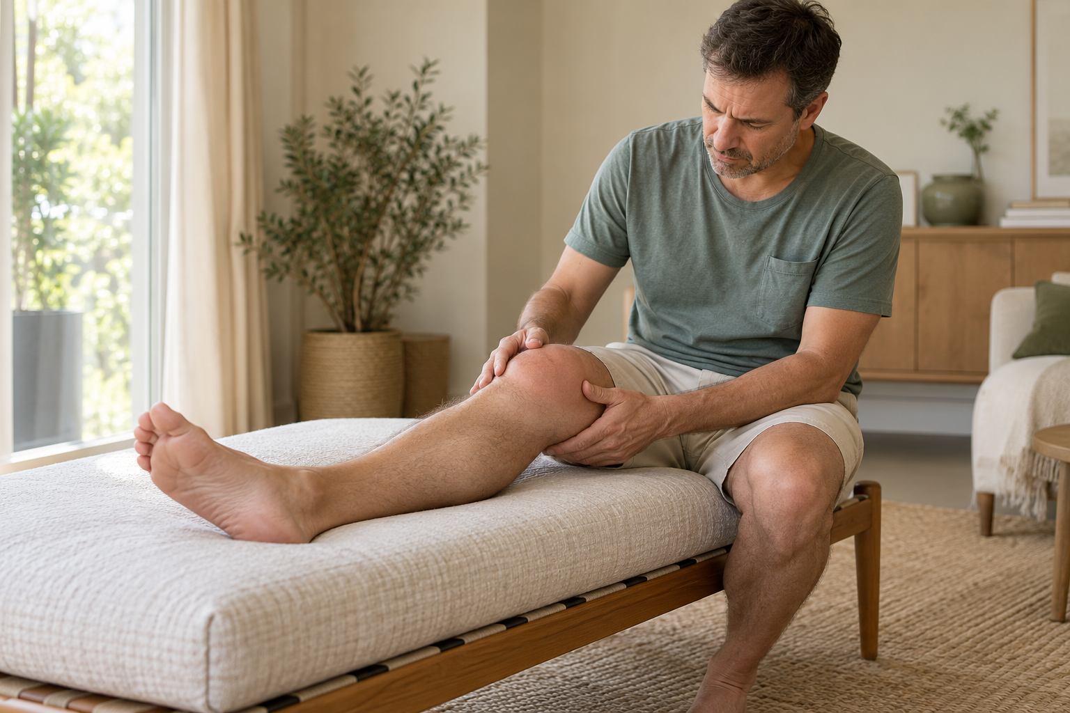
After an injury, symptoms usually appear quickly. Their intensity depends on the type of fracture and any associated injuries.
- Significant pain in the knee or thigh. It can increase when the leg moves or when attempting to put weight on it.
- Swelling. The knee can swell rapidly after the injury.
- Difficulty walking or moving the leg. The loading process can be painful, or even impossible.
- Deformity or bruise. A change in the appearance of the leg or a bruise may appear, but their absence does not rule out a fracture.
These signs can also accompany other knee injuries. After a significant trauma, a prompt medical evaluation is necessary to confirm the diagnosis and assess the condition of the leg.
HOW DOES THIS FRACTURE OCCUR?
An intra-articular fracture of the femur most often occurs following trauma significant enough to reach the end of the femur and the surface of the knee. The mechanism of injury and any existing bone fragility influence the shape of the fracture.
Direct collision
A fall or a direct impact on the knee can transmit a significant force to the end of the femur.
High-energy trauma
A road accident or other violent trauma can cause a complex fracture, sometimes accompanied by other injuries.
Fall with twisting or compression
Depending on the position of the leg and the intensity of the impact, the force can be concentrated on the lower part of the femur.
weakened bone
In some people, bone fragility can increase the risk of fracture after a minor trauma.

MAKE THE RIGHT DIAGNOSIS
The diagnosis is based on the circumstances of the trauma, examination of the leg, and imaging. The latter helps to determine the path of the fracture and its extension to the joint.
01 — Assess the leg and knee
The doctor looks for pain, swelling, any possible deformity and checks circulation as well as the sensitivity of the foot.
02 — Performing X-rays
X-rays generally allow identification of the fracture and assessment of its displacement.
03 — Specify the joint involvement
A CT scan may be requested to better examine the fragments and the joint surface, particularly to prepare for treatment. It is not a replacement for routine MRI.
The choice of treatment depends on all clinical and radiological findings.
PROTECT THE KNEE AND SUPPORT RECOVERY
Non-surgical treatment may be considered in certain stable, minimally displaced or non-displaced fractures, when alignment and the articular surface can be maintained without intervention. The decision rests with the medical team after reviewing the examinations.
The treatment aims to protect the fracture, control pain and gradually preserve mobility, while monitoring consolidation and alignment.
Protection and immobilization
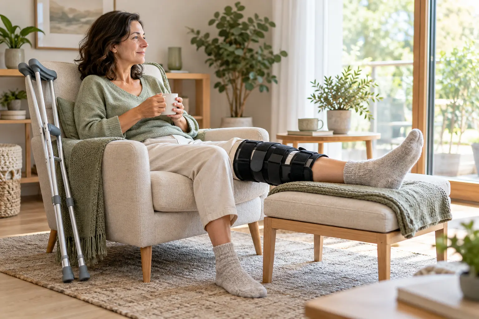
A splint or other protective device may be prescribed to limit movements that could put stress on the fracture. The duration and method are determined by the medical team based on the stability of the injury.
Support and movement
Weight-bearing on the leg may need to be limited or prohibited initially. Crutches can assist with mobility, following the doctor's instructions.
Pain and monitoring
Pain management is tailored to the individual's medical condition. Clinical and radiological checks are performed to ensure that the fracture remains stable and heals favorably.
Mobility and physiotherapy
When permitted, mobilization and physiotherapy are gradually resumed to maintain knee mobility and function. Exercises and their progression must adhere to the guidelines related to the fracture.
Protection and resumption of movement must be adapted to the stability of the fracture and its evolution.
WHEN DOES INTERVENTION BECOME NECESSARY?
A displaced or unstable intra-articular fracture may require intervention to restore alignment and the articular surface. The choice of procedure depends on the shape of the fracture, the condition of the tissues, and the individual's medical history.
Surgery is not automatic for every femur fracture. It is considered when the expected benefits of stabilization outweigh the risks, after analysis of examinations and the patient's general condition.
Displaced articular surface
When the fragments have shifted and deformed the surface of the knee, intervention may be discussed to try to restore proper alignment.
Unstable or complex fracture
Multiple fragments, an unstable fracture, or difficulty maintaining bone position may lead to consideration of surgical fixation.
Decision tailored to each situation
Age, bone quality, associated lesions, general condition and functional goals are taken into account in the choice of treatment.
RESUME WALKING AND ACTIVITIES GRADUALLY
After an intra-articular fracture of the femur, the resumption of movement and weight-bearing must follow the instructions of the medical team. Weight-bearing too early can compromise the stability of certain fractures.
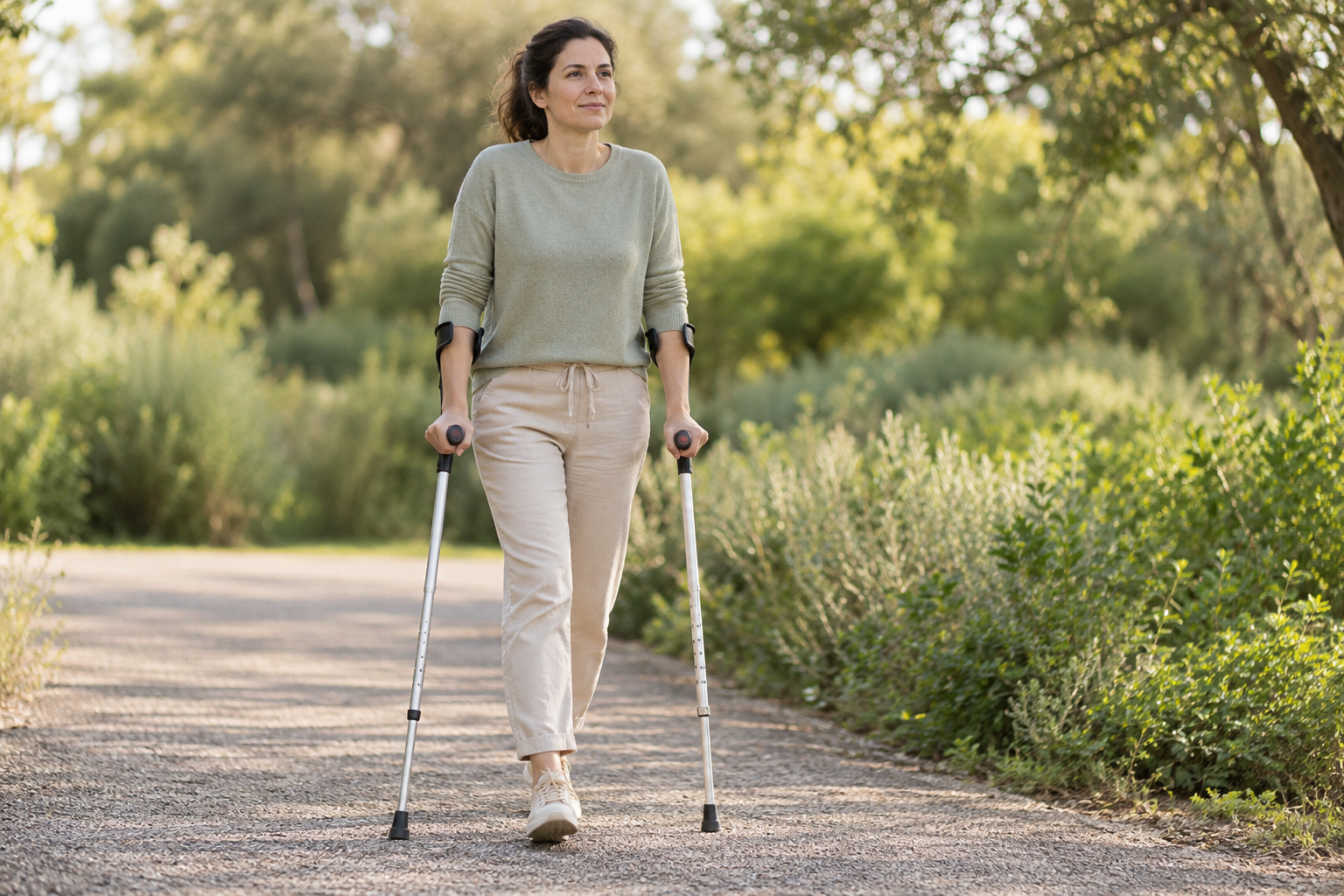
Initially, walking may require crutches and knee protection. Mobility and physiotherapy are introduced gradually as permitted.
Any increasing pain or swelling should be reported to the healthcare team. Exercises and movement patterns are adjusted according to the symptoms and monitoring of the fracture.
Walking, daily activities, and then more demanding activities are resumed in stages, depending on the consolidation, mobility, and strength regained.
Progress is made according to the evolution of the fracture and medical recommendations.

A CARE APPROACH TAILORED TO EACH PATIENT
An intra-articular fracture of the femur is not treated the same way in everyone. The displacement of the fragments, the extent of joint damage, and associated injuries influence the possible treatment options.
Dr. Benoît Coulin takes into account symptoms, mobility, usual activities, general condition, bone quality and the results of clinical examination and imaging.
Conservative or surgical treatment is discussed based on the stability of the fracture and the individual's needs. The objectives are explained to the patient, along with the protective and functional recovery steps tailored to their specific case.
«"Each fracture must be assessed according to its shape, stability, and the individual's situation."»
FREQUENTLY ASKED QUESTIONS
Have you suffered a knee or thigh injury?
A clinical examination, combined if necessary with appropriate imaging, helps to determine the origin of the symptoms and to propose a personalized treatment plan.
