

TIBIAL PLATFORM FRACTURE
The tibial plateau is the upper part of the tibia that forms part of the knee joint. A fracture in this area can be accompanied by displacement or depression of the bone, and sometimes by damage to the menisci or ligaments.
Treatment depends on the type and stability of the fracture. The goal is to protect the joint during healing and to choose a treatment tailored to each patient's individual situation.
UNDERSTANDING TIBIAL PLATFORM FRACTURE

-
This bony surface is located on the inner side of the knee and receives part of the femur.
-
Located on the outside, it can be touched alone or with the inner tray.
-
Covered with cartilage, it allows movement between the femur and the tibia. A fracture can crack or depress it.
-
The meniscus is located between the femur and the tibia. It can be injured during the same trauma.
RECOGNIZING THE SYMPTOMS
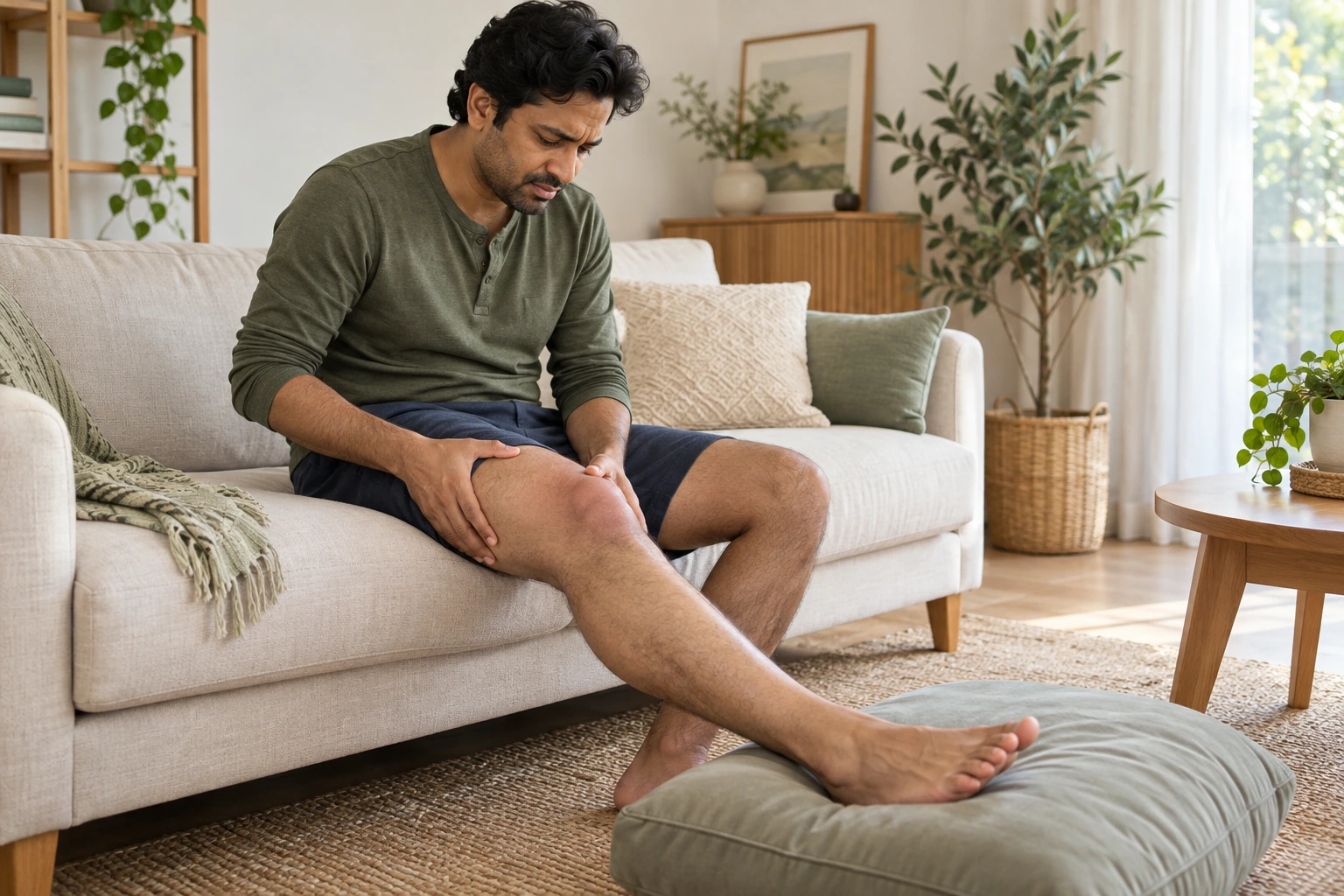
Symptoms appear after trauma, such as a fall, impact, or accident. Their intensity varies depending on the fracture and any associated injuries.
- Knee pain: It is often pronounced and worsens when the leg is used. Walking can become difficult.
- Swelling : The knee can swell rapidly after the injury. The swelling can limit movement.
- Difficulty gaining support: Weight-bearing may be painful, very limited, or impossible. Do not attempt to walk to "test" the knee.
- Reduced mobility: Bending or straightening the knee may become difficult. A feeling of instability or deformity may also be present.
These signs can have other causes and are not sufficient on their own to confirm a fracture. Significant pain, rapid swelling, or the inability to bear weight after an injury warrants urgent evaluation.
→ Consult the emergency signs of a tibial plateau fracture
HOW DOES THIS FRACTURE OCCUR?
A tibial plateau fracture is usually caused by trauma that transmits a significant force to the knee. The direction of the impact, its intensity, and the quality of the bone influence the shape of the fracture.
A fall or a sudden landing
A fall or landing with an impact on the leg can compress the upper part of the tibia and fracture its articular surface.
Direct collision
A significant impact to the knee, particularly during an accident, can cause a fracture of varying extent.
weakened bone
Bone fragility can increase the risk of fracture after a minor injury. It is taken into account in the overall assessment.
Associated lesions
The trauma can also affect the menisci, ligaments, or cartilage. Identifying these structures helps to determine the appropriate treatment.

MAKE THE RIGHT DIAGNOSIS
The diagnosis is based on the circumstances of the trauma, the clinical examination, and appropriate imaging studies. A CT scan can clarify the shape of the fracture and any depression; an MRI is reserved for situations where certain associated structures need to be examined.
01 — THE CIRCUMSTANCES OF THE TRAUMA
The doctor asks how the injury occurred, when the pain started, and if weight-bearing is possible.
02 — THE CLINICAL EXAMINATION
The examination assesses swelling, tenderness, mobility, and the condition of circulation and sensation in the foot, taking pain into account.
03 — IMAGING EXAMS
X-rays are used to look for the fracture. A CT scan often clarifies the displacement and depression; an MRI may be recommended if damage to the meniscus or ligaments is suspected.
An image must be interpreted in conjunction with the clinical examination and symptoms to guide management.
TREATING FRACTURE WITHOUT SURGERY
Some stable fractures with little or no displacement can be treated without surgery. Management then relies on protecting the knee, specific weight-bearing instructions, and medical follow-up.
The goal is to allow the bone to consolidate in a satisfactory position, while progressively preserving the mobility and function of the knee.
PROTECT THE LEG
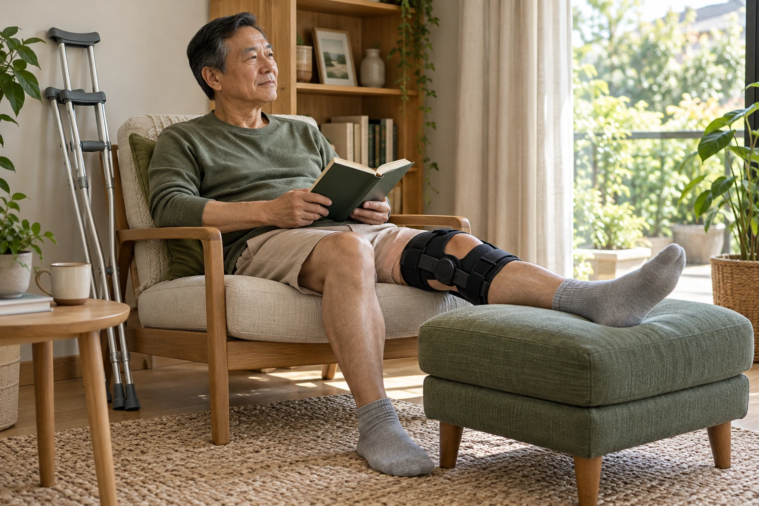
Depending on the fracture, weight-bearing may be prohibited or limited for a period determined by the medical team. Crutches can help with this. Weight-bearing should not be resumed without authorization.
SPLINTING AND IMMOBILIZATION
A brace may be prescribed when it is useful to protect the knee. The duration of use depends on the stability of the fracture and medical instructions.
MONITOR THE CONSOLIDATION
Clinical and radiological examinations allow monitoring of the fracture's progress. Any worsening of pain, swelling, or new difficulties should be reported to the healthcare team.
GRADUALLY RESUME MOBILITY
When the fracture allows, mobility and physiotherapy are gradually reintroduced. Exercises must adhere to weight-bearing and protective guidelines.
Fracture protection and adherence to weight-bearing guidelines accompany each stage of recovery.
IN WHAT SITUATIONS SHOULD WE OPERATE?
Surgery may be considered if the fracture is displaced, unstable, or if the impaction compromises the articular surface. The decision also depends on the tissues around the knee, any associated injuries, and the patient's overall condition.
Surgery generally aims to reposition and stabilize the fragments. The timing and technique of the surgery depend on the fracture and the condition of the tissues.
Displaced fracture
When bone fragments are displaced, intervention may be discussed to restore satisfactory alignment and stabilize the fracture.
Depression of the articular surface
Significant depression of the endplate can alter the joint surface. A CT scan helps assess the fracture and discuss treatment options.
Unstable or complex fracture
Some fractures require surgical stabilization. Associated injuries or the condition of the tissues may alter the strategy and timing.
RESUME ACTIVITIES GRADUALLY
Initially, the priority is to protect the fracture. Support and mobilization must follow the instructions of the medical team.
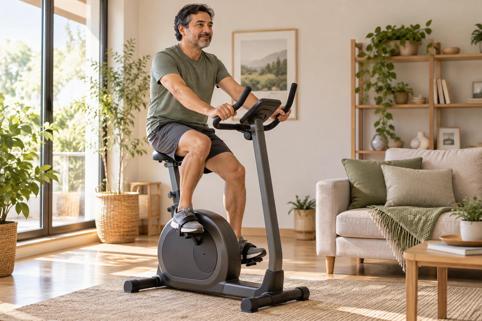
When the bone has healed sufficiently, physiotherapy can help gradually restore knee mobility and muscle strength. Exercises are adjusted according to the progress of the fracture.
Walking and daily activities can resume depending on medical clearance and the knee's ability to withstand stress. Significantly increasing pain or worsening swelling should prompt a reassessment of activity with the healthcare team.
Sports activities can be resumed later, after assessment of the healing, mobility, strength, and control of the knee. The timeline varies depending on the fracture, the treatment performed, and any associated injuries.
Progress depends on the observed healing and medical instructions, not on a uniform timeframe for everyone.
→ Discover the steps of knee rehabilitation

A CARE APPROACH TAILORED TO EACH PATIENT
There is no single treatment for all tibial plateau fractures. Two fractures in the same location can differ in their stability, displacement, or the structures of the knee they affect.
Dr. Benoît Coulin considers the patient's symptoms, clinical examination, imaging, associated injuries, and functional needs. Age, activities, occupation, and recovery goals are also taken into account in the discussion.
Depending on the situation, treatment may involve protection and monitoring, or require surgical stabilization. The potential benefits, limitations, and recovery stages are explained to the patient.
«"The right decision begins with a precise understanding of the fracture and its consequences for the patient."»
FREQUENTLY ASKED QUESTIONS
IS YOUR KNEE PAINFUL AFTER A TRAUMA?
A clinical examination and appropriate imaging will determine the nature of the injury and the treatment options. In case of significant pain or inability to bear weight, request an urgent evaluation.
