

calcaneus fracture
A calcaneus fracture affects the heel bone, which bears part of the body's weight and participates in hindfoot movement. When it is displaced or involves the subtalar joint, surgery may sometimes be recommended to reposition and stabilize the fragments.
The decision depends on the clinical examination, imaging, the condition of the skin and tissues around the heel, any associated injuries, the patient's overall health, and their goals. A calcaneal fracture does not always require surgery.
UNDERSTANDING SURGERY FOR A MEDIAL MALLEOLAR FRACTURE

-
This bony surface is located on the inner side of the knee and receives part of the femur.
-
Located on the outside, it can be touched alone or with the inner tray.
-
Covered with cartilage, it allows movement between the femur and the tibia. A fracture can crack or depress it.
-
The meniscus is located between the femur and the tibia. It can be injured during the same trauma.
WHEN SHOULD SURGERY BE CONSIDERED FOR A CALCANEUS FRACTURE?
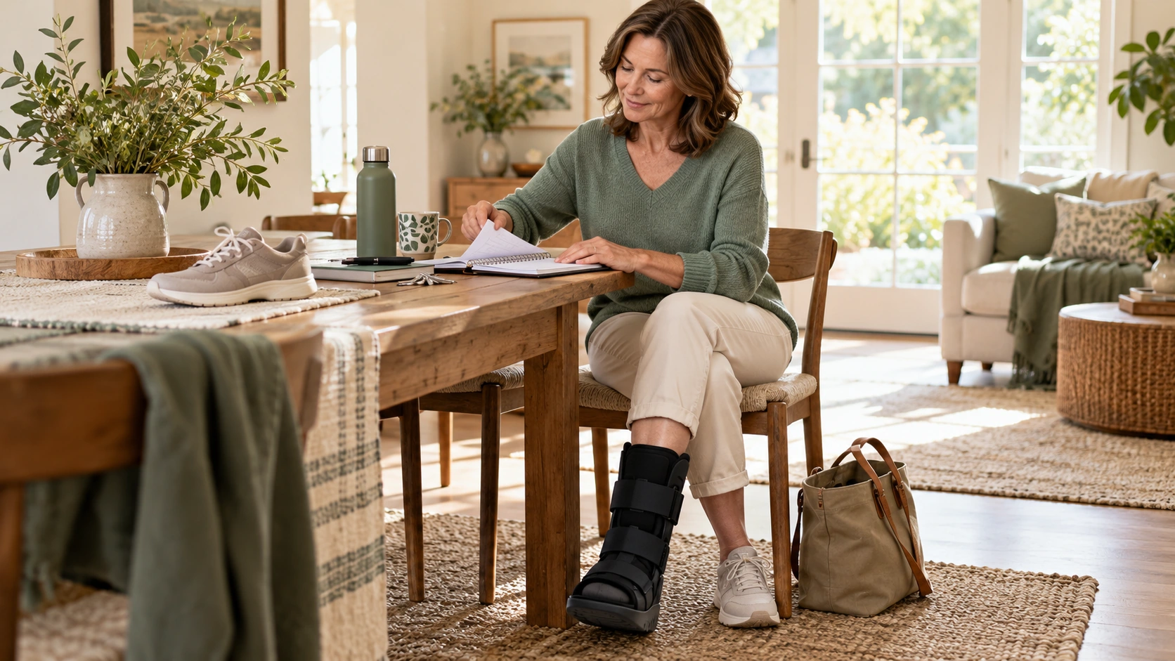
Surgery may be considered when the shape and displacement of the fracture compromise heel alignment or the joint surface. The decision is based on X-rays and often a CT scan, as well as examination of the foot and skin. Minimally displaced fractures are often treated non-operatively.
- Significant displacement of fragments — Repositioning may be considered if the displacement alters the shape or alignment of the calcaneus.
- Displaced joint injury — A fracture that alters the subtalar surface may justify a reduction in order to restore its congruity as much as possible.
- Heel deformity — A significant loss of height, widening or poor alignment of the rear foot can weigh in the decision.
- Fragment threatening the skin or tendon insertion involved — Certain specific fractures, particularly those with a displaced posterior fragment, require rapid assessment.
The procedure is not automatic. The condition of the skin, swelling, other injuries, the risks associated with anesthesia, and the expected benefits are also taken into account. An abnormal image alone is not sufficient to make a decision. The strategy may include non-surgical treatment and monitoring. In cases of recent foot trauma, see the page on the Coulin Center of Movement in Geneva presents orthopedic and trauma care.
PREPARE FOR THE INTERVENTION
The preparation aims to determine the fracture and to choose the appropriate timing and technique. Swelling and the condition of the heel tissues are particularly important for planning the intervention.
Preoperative assessment
The team reviews the examinations, assesses the skin, circulation, and sensation of the foot, and looks for any associated injuries. An anesthesia consultation and, depending on the situation, further tests or examinations are arranged.
Intervention planning
X-rays and CT scans help to determine the number and position of the fragments, as well as the extent of joint involvement. This information guides the choice of surgical approach and fixation material.
Physical preparation and practice
Since support is often limited after surgery, it is helpful to arrange for home visits and necessary assistance. Training in the use of crutches may be offered depending on the situation.
Instructions before the operation
The team will provide instructions regarding fasting, hygiene, and admission. Do not change or stop any medication without consulting the medical team.
HOW IS SURGERY FOR A CALCANEAL FRACTURE PERFORMED?
The procedure depends on the type of fracture, its displacement, the condition of the tissues, and the technique used. The surgeon verifies the position of the fragments using intraoperative imaging when necessary.
Installation and anesthesia
The patient is positioned to allow access to the heel. General anesthesia or another anesthetic strategy may be used depending on the anesthesiologist's assessment and the situation. After sterile preparation, the team verifies the side to be operated on and the patient's medical record.
Access to the treated area
The surgeon chooses an incision appropriate to the fracture. Depending on the case, access may be wider, more extensive, or percutaneous. The choice must allow for manipulation of the fragments while taking into account the delicate skin covering the heel.
Main surgical procedure
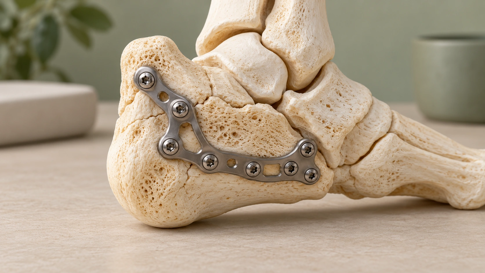
The fragments are mobilized and repositioned to restore as much as possible the shape of the calcaneus, its alignment, and, if affected, the surface of the subtalar joint. Instruments and radiographic guidance may be used to assist in the maneuvers. Once reduction is achieved, the fragments are stabilized with screws, sometimes with a plate. The exact hardware depends on the fracture configuration and the quality of the bone. The surgeon also verifies that the fixation does not interfere with surrounding structures.
result check
The team checks the position of the fragments, the alignment of the heel, and the position of the screws or plate. Intraoperative images can confirm the reduction and fixation. The check also takes into account the tissues around the incision.
Closure and end of the intervention
After verification, the tissues are closed and a dressing is applied. Immobilization may be implemented. The patient is then transferred to the recovery room for monitoring.
The priority is to choose a fixation and approach adapted to the fracture and the tissues of the heel.
The duration of the operation, the type of anesthesia, and the need for hospitalization vary depending on the complexity of the fracture and the patient's overall health. These factors are explained before the procedure.
WHICH SURGICAL TECHNIQUE SHOULD BE CHOOSED FOR A CALCANEUS FRACTURE?
Open reduction with internal fixation allows visualization and repositioning of fragments when a displaced fracture requires it. It may involve a plate and screws, but the condition of the heel tissues is a major factor in determining the timing and approach.
Less invasive techniques can be considered in certain situations. The choice is made on a case-by-case basis: each method has its indications, advantages, and limitations. The strategy is guided by the anatomy of the fracture and the risks of skin complications.
Discount open
An incision allows access to the fragments and their repositioning under direct control. This approach can be useful for complex fractures, but it puts more stress on the tissues and requires careful attention to wound healing.
Less invasive or percutaneous approach
Smaller incisions and specialized instruments allow for the manipulation and fixation of certain fragments. This option depends on the fracture configuration and the possibility of achieving satisfactory reduction.
Fixing by screws or plate and screws
Screws can stabilize the fragments; a plate can be added when the fracture geometry requires it. The hardware is chosen according to the bone, the fracture, and the reduction achieved.
Timing of the intervention and condition of the tissues
When swelling is significant, surgery may be postponed until the tissues are more favorable, if the situation allows. In cases of open fracture or skin injury, management is tailored to the urgency and the extent of the damage.
The chosen technique aims for appropriate fixation, while taking into account the skin, the joint and the functional objectives.
AFTER THE PROCEDURE: THE FIRST STEPS
After the operation, the team monitors the pain, the dressing, and the condition of the foot. Instructions for weight-bearing and immobilization are given based on the fixation and checks performed.
Recovery room and pain
Monitoring focuses on pain, sensitivity, and circulation in the foot. The team implements appropriate pain management and explains how to take the prescribed medication.
Support, walking and immobilization
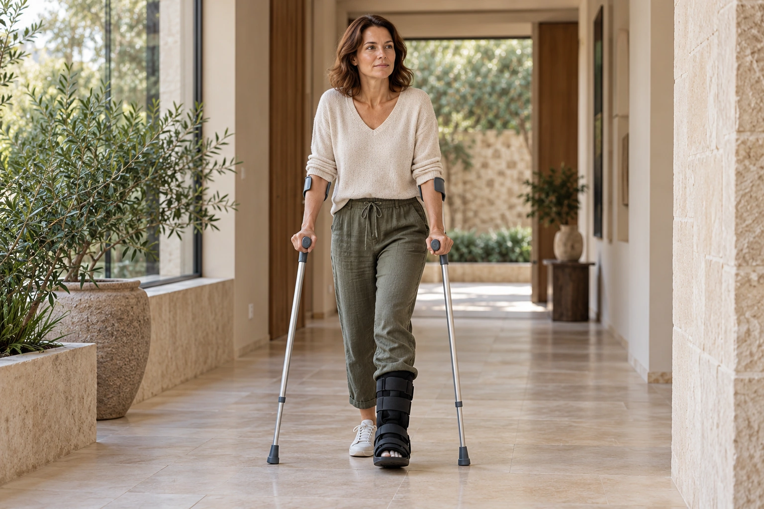
Weight-bearing is generally prohibited or limited initially, but the specific instructions depend on the fracture and its fixation. Crutches are often necessary. A splint or boot may be recommended. It is essential to follow the team's precise instructions and not resume weight-bearing without approval.
Return home and monitoring
The length of hospital stay depends on the medical situation, pain management, and the patient's level of independence. The team will explain dressing changes, wound monitoring, and the follow-up appointment. Contact a healthcare professional promptly if the pain increases significantly, if the wound becomes red or oozes, if you develop a fever, or if your foot becomes cold, pale, numb, or unusually painful.
REHABILITATION AND RETURN TO ACTIVITIES AFTER A CALCANEUS FRACTURE
The initial steps focus on protecting the fixation, monitoring healing, and controlling pain and swelling. Permitted ankle and foot movements depend on the surgeon's instructions and the condition of the tissues.
Rehabilitation progresses according to consolidation and checks. It can work on mobility, strength, balance and gait quality, without exceeding the weight-bearing limits defined by the team.
Resuming walking, climbing stairs, driving, and work depends on the affected foot, the level of pain, the permitted weight-bearing, and the demands of the activity. Temporary adjustments may be necessary, especially for physically demanding jobs.
Sports activities resume gradually, generally after medical clearance and when the body's strength, mobility, and balance allow. Activities involving impact or changes of direction require specific assessment.
Progress is made according to consolidation and regained capabilities, without a guaranteed timetable. Physiotherapy and rehabilitation can accompany this recovery when recommended by the team.

EXPECTED RESULTS, LIMITATIONS AND RISKS
Objectives and expected results
The surgery aims to restore the shape of the heel, its alignment, and, if applicable, joint congruity as much as possible. It also aims to stabilize the fragments to allow them to heal.
Boundaries
The outcome depends on the severity of the fracture, the extent of cartilage damage, the condition of the tissues, and the healing process. Stiffness, pain, or difficulty walking may persist. Subtalar osteoarthritis can develop after certain joint fractures, even when treatment is appropriate.
Risks and complications
Possible complications include impaired wound healing or infection, hematoma, phlebitis, stiffness, or persistent pain. Malunion, poor bone healing, or fixation problems may sometimes require additional treatment.
Surgery aims to improve alignment and stability, but cannot guarantee the absence of pain or after-effects.

A CARE APPROACH TAILORED TO EACH PATIENT
Two people with similar calcaneal fractures on imaging do not necessarily require the same intervention. The condition of the skin, the degree of displacement, and the functional goals can lead to different strategies.
Dr. Benoît Coulin takes into account age, anatomy, bone and tissue quality, the severity of the fracture, associated injuries, activity level, occupation, previous treatments, and personal goals. These factors help to weigh the expected benefits against the risks.
The technique, timing of the intervention, and recovery program are tailored to the fracture and the patient's individual situation. The decision is explained during a consultation with the medical team. To learn more about the Dr. Benoît Coulin, see the center's page.
«"The surgical strategy is developed based on the fracture, the condition of the tissues, and the functional needs of each patient."»
FREQUENTLY ASKED QUESTIONS
Is surgery indicated for your calcaneus fracture?
A clinical examination and imaging analysis allow us to assess the fracture and discuss the most appropriate strategy for your situation.
