

fracture of the medial malleolus
A fracture of the medial malleolus affects the bony prominence located inside the ankle, at the end of the tibia. When the fragments are displaced or the joint is unstable, surgery may be considered to realign and stabilize them using appropriate hardware.
Surgery is not always necessary. The decision takes into account the displacement, ankle stability, any other fractures or associated injuries, the clinical examination, and the patient's overall condition. An isolated, stable, and minimally displaced fracture can sometimes be treated without surgery.
UNDERSTANDING SURGERY FOR A MEDIAL MALLEOLAR FRACTURE

-
This bony surface is located on the inner side of the knee and receives part of the femur.
-
Located on the outside, it can be touched alone or with the inner tray.
-
Covered with cartilage, it allows movement between the femur and the tibia. A fracture can crack or depress it.
-
The meniscus is located between the femur and the tibia. It can be injured during the same trauma.

WHEN SHOULD SURGERY BE CONSIDERED FOR A MEDIAL MALLEOLUS FRACTURE?
Surgery may be recommended when maintaining the fragments in the correct position is necessary to restore ankle stability. The decision is based on an analysis of the fracture as a whole, not solely on the results of an image.
Displaced fracture
If the bone fragments are separated or misaligned, reduction and fixation may be discussed.
Unstable ankle
A fracture associated with instability of the ankle mortise may require surgical stabilization.
Complex or associated fracture
Involvement of multiple malleoli, the syndesmosis, or other structures may alter the strategy and lead to treating multiple areas.
Insufficient reduction or difficulty in maintaining
An intervention may be considered if alignment cannot be achieved or maintained by non-surgical treatment.
An isolated, stable, and minimally displaced fracture does not always require surgery. The choice also depends on the skin, vascular and neurological status, medical history, activities, and individual risk factors. To learn more about orthopedic and trauma care in Geneva, consult the [website/link]. Coulin Center of Movement.
PREPARE FOR THE INTERVENTION
The preparation depends on the type of fracture, the condition of the ankle, and when the procedure can be performed. Swelling and the condition of the skin are among the factors taken into account.
Preoperative assessment
Clinical examination and X-rays allow for the assessment of the fracture. A CT scan may be requested to determine the shape of the fragments or the extent of joint surface involvement. An anesthesia consultation and, depending on the situation, further tests complete the evaluation.
Intervention planning
The surgeon analyzes the alignment, size, and orientation of the fragments, as well as any associated fractures. These elements guide the choice of surgical approach and fixation hardware.
Physical preparation and practice
It may be helpful to arrange for the return home and for getting around with crutches. The team will specify the necessary assistance based on the support guidelines provided after the procedure.
Instructions before the operation
Fasting, hygiene, and arrival instructions will be provided by the medical team. Do not change or stop any medication without consulting your healthcare professionals.

HOW DOES THE PROCEDURE WORK?
The technique depends on the fracture configuration and associated injuries. The goal is to restore ankle alignment and stabilize the fragments that need to be fixed.
Installation and anesthesia
The procedure is performed under general or regional anesthesia, depending on the anesthetic assessment and the clinical situation. The skin is prepared under sterile conditions, and the team reviews the side to be operated on and any available images.
Access to the treated area
A brace may be prescribed when it is useful to protect the knee. The duration of use depends on the stability of the fracture and medical instructions.
Reduction and fixation of the fracture
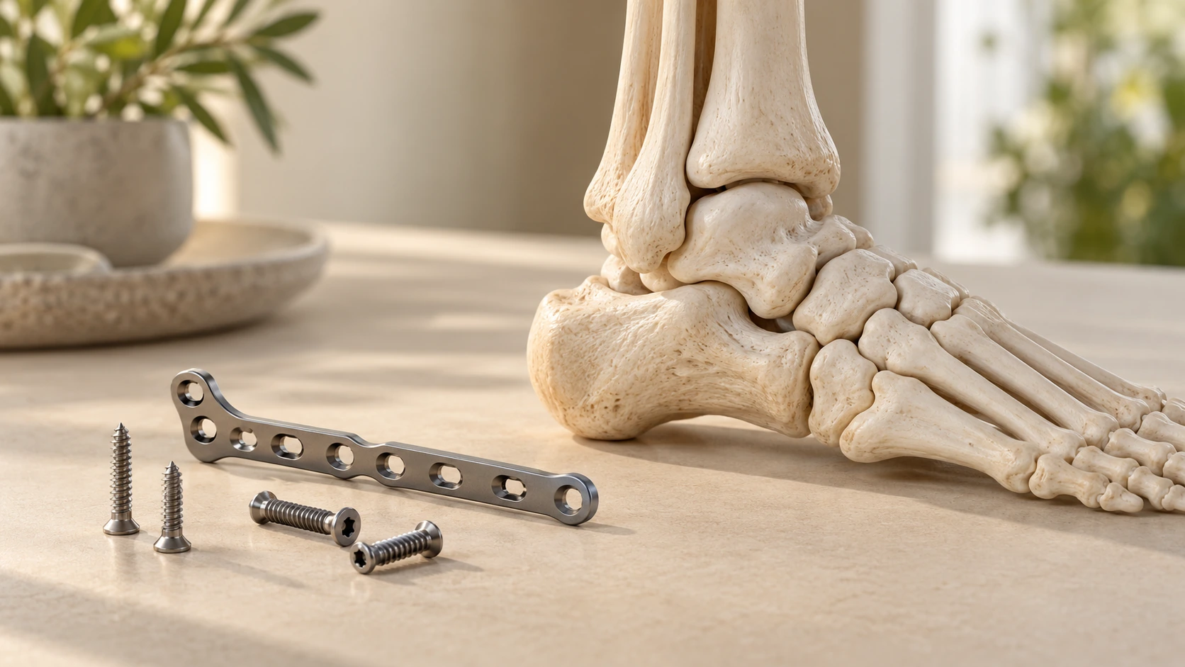
The fragments are repositioned under direct visual control. The surgeon removes any tissue that might be hindering reduction, if necessary, and then checks the joint's congruity. Fixation is tailored to the size, shape, and quality of the fragments: it may involve one or more screws, a plate, or another fixation device. If other fractures are causing ankle instability, they can be addressed during the same procedure. The goal is to maintain reduction during consolidation, without imposing an identical solution on all patients.
result check
The alignment and position of the hardware are monitored during the procedure, including intraoperative X-ray imaging if necessary. The surgeon verifies that the fixation is stable and that the hardware does not interfere with the joint.
Closure and end of the intervention
After the checks are performed, the tissues are closed and a dressing is applied. A splint or immobilization device may be put in place depending on the stability achieved and the protective measures in place. The patient is then transferred to the recovery room.
The fixation must maintain a satisfactory reduction while respecting the anatomy of the ankle.
The duration of the procedure, the type of anesthesia and the method of hospitalization vary depending on the complexity of the fracture, the associated procedures and the organization of the facility.
WHICH SURGICAL TECHNIQUE SHOULD BE CHOOSED?
The choice of fixation depends on the shape of the fracture, the size of the fragments, the bone quality, and the stability of the ankle. Associated fractures may require a more extensive surgical plan.
Anatomical reduction and stable fixation are the goal when surgery is indicated. The options described below are not interchangeable in all situations.
Screw fixing
One or more screws can hold a sufficiently large fragment in place after reduction. Their direction is chosen according to the fracture configuration. This option is not suitable for all fragments, particularly when they are very small or fragmented.
Plate and screws
A plate may be used when the morphology of the fracture or the desired stability warrants it. It allows for the retention of several fragments, but the choice depends on the available space and the quality of the tissues.
Strapping or guy wire assembly
In some fractures, a cable-link fixation can help stabilize the fragment. It is chosen based on the shape of the fracture and the mechanical stresses to be controlled.
Treatment of associated fractures
When the medial malleolus is part of a bimalleolar or trimalleolar fracture, or when syndesmosis instability is identified, further procedures may be necessary. The order and method of fixation are determined based on a complete assessment of the ankle.
The technique is chosen according to the fracture and the stabilization objectives, after analysis of the images and clinical examination.
AFTER THE PROCEDURE: THE FIRST STEPS
After the operation, the team monitors pain, the dressing, circulation, and sensation in the foot. The length of stay depends on the procedure, the patient's overall condition, and the conditions for returning home.
Recovery room and pain
Pain is assessed and managed according to the team's instructions. Monitoring also includes checking the dressing and toe sensitivity.
Support, walking and immobilization
Weight-bearing may be prohibited, partial, or permitted depending on the fracture and the stability of the fixation. Crutches are often helpful initially. A splint or boot may be prescribed; the exact instructions will be given by the medical team, who will also indicate when to mobilize the ankle.
Return home and monitoring
The patient may be discharged the same day or after a short hospital stay, depending on the situation. They will receive instructions for dressing changes, leg elevation, and follow-up care. Significantly worsening pain, fever, wound drainage, or cold, pale, or numb toes warrant prompt contact with the medical team.
REHABILITATION AND RESUMPTION OF ACTIVITIES AFTER A MEDIAL MALLEOLAR FRACTURE
Initially, the goals are to protect the fixation, monitor healing, and limit pain and swelling. Elevation of the limb and permitted toe movements may be part of the instructions.
Rehabilitation progresses according to the consolidation of the injury and the surgeon's instructions. It can then focus on ankle mobility, strength, balance, and movement control, with exercises adapted to the stage of recovery.
Walking, climbing stairs, driving, and returning to work depend on support guidelines, mobility, and the ability to move safely. Physical work activities may require a different progression than sedentary tasks.
The return to sport is gradual and depends on the consolidation of injuries, mobility, strength, and balance. It takes place after medical clearance, taking into account the activity practiced and the patient's progress.
The resumption of activities is built step by step, according to consolidation and functional recovery.

EXPECTED RESULTS, LIMITATIONS AND RISKS
Objectives and expected results
Surgery aims to realign the fragments and stabilize the ankle when the fracture requires it. It seeks to create conditions favorable to healing and the recovery of function.
Boundaries
Healing and recovery vary depending on the fracture, associated injuries, the patient's overall health, and mechanical stresses. Stiffness, discomfort, or pain may persist. Fixation does not guarantee the same recovery for all patients.
Risks and complications
Like any surgical procedure, this surgery carries risks, including infection, hematoma, wound healing problems, phlebitis, stiffness, or persistent pain. More rarely, a problem with fixation or consolidation may necessitate further treatment or a second surgery.
The expected benefit of stabilization is weighed against the limitations of the fracture and the risks inherent in the intervention.

A CARE APPROACH TAILORED TO EACH PATIENT
Two people with a similar-looking medial malleolar fracture may have different needs. Displacement, ankle stability, and associated injuries influence treatment.
Dr. Benoît Coulin takes into account age, anatomy, bone and skin quality, the severity of the fracture, activity level, occupation, medical history, and personal goals. General health and current treatments are also considered in the preparation.
The technique, the timing of the procedure, the fixation materials, and the recovery program are all tailored to these factors. The options and their limitations are explained to the patient to enable them to make an informed decision.
«"The choice of treatment depends on the stability of the ankle, the fracture, and each patient's individual situation."»
FREQUENTLY ASKED QUESTIONS
Does a fracture of the medial malleolus require surgery?
A clinical examination and analysis of radiographs, sometimes supplemented by a scan, allow for the assessment of ankle stability and discussion of the appropriate treatment.
