

Fracture of the lateral malleolus
A lateral malleolus fracture affects the lower end of the fibula, the bone located on the outer side of the leg. It often occurs after an ankle sprain or a fall. Treatment aims to allow the bone to heal and to preserve the alignment and stability of the joint.
A fracture of the lateral malleolus does not always require surgery. The decision depends in particular on the displacement of the fragments, the stability of the ankle, any associated injuries, the clinical examination, and the patient's needs.
UNDERSTANDING SURGERY FOR A MEDIAL MALLEOLAR FRACTURE

-
This bony surface is located on the inner side of the knee and receives part of the femur.
-
Located on the outside, it can be touched alone or with the inner tray.
-
Covered with cartilage, it allows movement between the femur and the tibia. A fracture can crack or depress it.
-
The meniscus is located between the femur and the tibia. It can be injured during the same trauma.
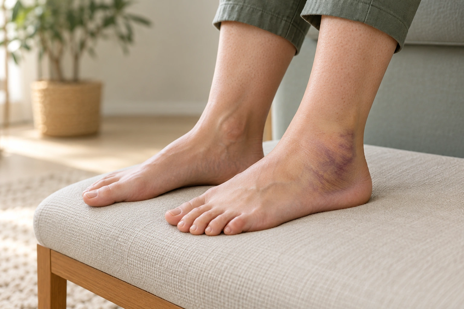
RECOGNISING THE SIGNS OF A LATERAL MALLEOLUS FRACTURE
After a twist or impact, a fracture can cause pain on the outer side of the ankle. The signs vary depending on the severity of the injury.
- Localized pain around the lateral malleolus, enlarged by movement or pressure.
- Swelling and bruising which can appear quickly around the ankle and foot.
- Difficulty gaining support or to walk normally.
- Touch sensitivity at the level of the bone, sometimes associated with a feeling of instability.
A deformity, a wound, a cold or numb foot, or significant pain warrants prompt medical evaluation. After an acute injury, you can consult the SOS Knee Geneva orthopedic emergency service, which indicates that it takes care of ankle and foot fractures.
HOW DOES THE FRACTURE OCCUR?
The mechanism helps to understand the fracture, but imaging and clinical examination are needed to determine its type and stability.
ANKLE TWIST
A foot that turns inwards or outwards can transmit a significant force to the fibula.
FALL OR FALSE STEP
A fall, a slip or a bad landing can cause a fracture, sometimes with other ankle injuries.
SPORTS TRAUMA
Changes of direction, contact and landings after a jump can result in a twist or impact.
BONE FRAGILITY
Bone fragility can increase the risk of fracture even from minor trauma. The patient's medical history is taken into account in the assessment.

ASSESSING THE ANKLE FRACTURE AND STABILITY
Clinical examination
The doctor examines the pain, swelling, skin, sensitivity, circulation of the foot and the ability to bear weight, when this can be assessed safely.
X-rays
X-rays allow the fracture to be located, the displacement to be assessed, and the joint alignment to be verified. Additional X-rays may be requested if stability remains uncertain.
Physical preparation and practice
It may be helpful to arrange for the return home and for getting around with crutches. The team will specify the necessary assistance based on the support guidelines provided after the procedure.
Further tests if necessary
A CT scan can be useful to clarify a complex fracture or to search for associated fragments. Imaging is chosen based on the results of the examination and X-rays.
The treatment decision depends on the stability of the ankle, not just on the presence of a fracture line.Reference sources emphasize the importance of verifying the retention of the talus in the mortise and the integrity of the medial and syndesmotic structures.
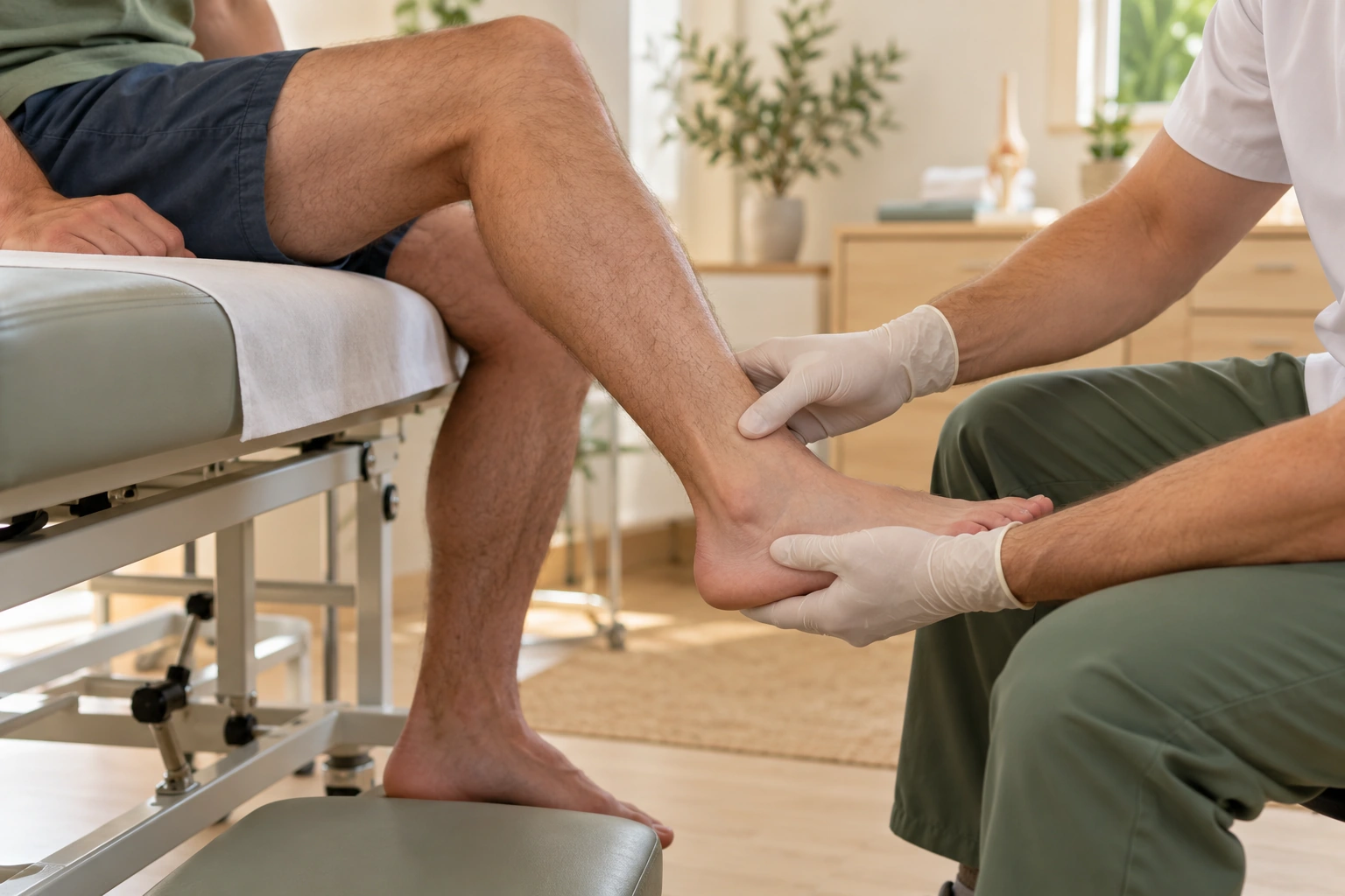
WHEN IS NON-SURGICAL TREATMENT CONSIDERED?
A stable, minimally displaced fracture can often be treated without surgery. Ankle protection, weight-bearing, and the resumption of movement are determined by the medical team based on the fracture and the examinations performed.
Protection and immobilization
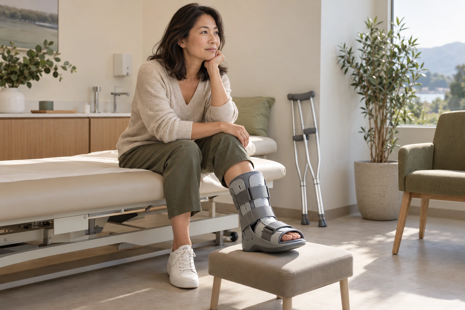
A boot or other form of immobilization may be recommended to protect the ankle during the healing process. The device and its duration depend on the type of fracture and its stability.
Support and movement
Weight-bearing may be permitted, limited, or temporarily avoided depending on medical instructions. Crutches can help ensure safe movement and adherence to the prescribed weight-bearing level.
Consolidation monitoring
Follow-up consultations and X-rays are used to verify that the fracture remains in satisfactory alignment. The frequency of these check-ups is tailored to each patient's individual case.
Mobility and rehabilitation
When the fracture allows, ankle movements and exercises are gradually reintroduced. Physiotherapy can help restore mobility, strength, and balance.
Stable fractures of the lateral malleolus are often managed without surgery, while displaced fractures or those associated with instability may require a different strategy.
WHEN TO CONSIDER SURGERY?
Surgery may be considered when the fracture compromises the alignment or stability of the ankle. The goal is to reposition the fragments correctly and hold them in place during the healing process.
Significant displacement
The fragments are no longer sufficiently aligned or risk losing their position.
Ankle instability
The talus is not properly centered in the mortise, or the ligaments and syndesmosis are affected.
Complex fracture or associated injuries
The fracture involves multiple fragments or is accompanied by a fracture of another malleolus or another injury requiring stabilization.
An isolated fracture of the lateral malleolus does not automatically lead to surgery. The decision takes into account all the images, the clinical examination, the condition of the skin, and the patient's characteristics.
HOW DOES THE PROCEDURE WORK?
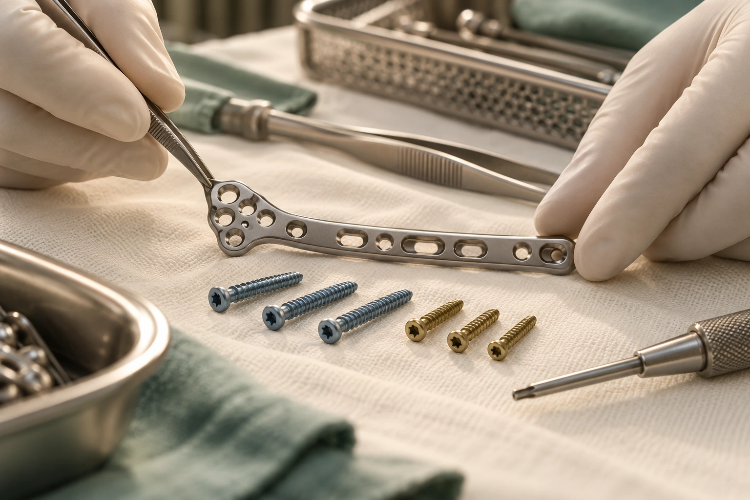
When surgery is indicated, it generally involves repositioning the fibula and stabilizing the fracture. The technique depends on the shape of the fracture, the bone quality, and any associated injuries.
INSTALLATION AND ANESTHESIA
The procedure takes place in the operating room, after verification of the side to be treated and the imaging file. General or regional anesthesia may be offered by the anesthesia team. The leg is positioned to allow access to the ankle and for X-ray examinations.
ACCESS TO THE FRACTURE
The surgeon accesses the fibula through an incision on the outer side of the ankle. He carefully exposes the area in order to reduce the fragments while preserving the surrounding tissues.
REDUCTION AND FIXING
The fragments are repositioned to restore the length, rotation, and alignment of the fibula. They are then held in place, often with a screw and plate adapted to the fracture. If syndesmosis involvement is suspected, its stability is assessed, and an additional device may be required.
ALIGNMENT CHECK
X-ray images taken during the procedure help to verify the position of the fragments, the alignment of the ankle, and the placement of the hardware. The surgeon also checks the stability of the joint and the relationship between the tibia and fibula.
CLOSURE AND END OF THE PROCEDURE
The wound is closed and protected with a dressing. A splint or boot may be applied depending on the situation. The patient is then transferred to the recovery room for monitoring.
The goal of fixation is to maintain the fracture in a position favorable to healing and to restore a stable ankle.
The duration of the procedure, the type of anesthesia, hospitalization, and postoperative support depend on the individual case and the hospital's protocol. Screw and plate fixation, as well as intraoperative radiological monitoring, are options described in surgical guidelines.
AFTER TREATMENT: THE FIRST STEPS
After a fracture of the lateral malleolus, the instructions are adapted to the treatment received, the stability of the ankle and the evolution of the fracture.
MONITORING AND PAIN
The team monitors pain, swelling, circulation, and sensitivity in the foot. Pain management is adjusted by the healthcare team.
SUPPORT, CRUTCHES AND IMMOBILIZATION
Weight-bearing may be permitted, partial, or delayed. Follow the instructions received and use crutches if prescribed. A boot or brace can protect the ankle.
RETURN HOME AND CHECKS
You may be able to return home the same day or after a short hospital stay, depending on the procedure and your medical condition. The dressing, wound care instructions, and follow-up appointment will be provided by the team. Contact a healthcare professional immediately if you experience significantly worsening pain, fever, a draining wound, or if your foot becomes cold, pale, or numb.
GRADUALLY REGAIN SUPPORT AND MOBILITY
Initially, the goals are to protect the fracture, limit pain and swelling, and monitor the healing process. The team explains leg positioning and comfort measures.
Resumption of ankle movement and walking depends on the stability of the fracture, the chosen treatment, and follow-up appointments. Physiotherapy can then focus on mobility, strength, balance, and weight-bearing.
Daily activities, climbing stairs, and driving are resumed gradually, depending on the ability to walk safely, the affected side, and medical instructions. Return to work depends on the job requirements and the healing of the fracture.
Sport is reintroduced gradually, after medical approval, when the patient's physical condition and functional abilities allow it. Activities involving jumping, changes of direction, or a risk of falling require a particularly cautious progression.
Recovery is built step by step, without an identical timetable for everyone. There physiotherapy and rehabilitation can be discussed with the team that is treating your fracture.


A CARE APPROACH TAILORED TO EACH PATIENT
Two fractures of the lateral malleolus that appear close together on an X-ray can have different consequences depending on their stability, displacement, and associated injuries.
Dr. Benoît Coulin assesses, among other things, the position of the fragments, the condition of the ligaments, the quality of the bone, the condition of the skin, the functional age, the patient's usual activities, and their goals. The opinion of Dr. Benoît Coulin may be asked to discuss diagnosis and treatment options.
Treatment, weight-bearing capacity, monitoring, and return to activities are determined based on the fracture and its progression. Intervention is recommended when the expected benefits outweigh the associated constraints and risks.
«"The choice of treatment is based on an analysis of the fracture, the stability of the ankle, and each patient's individual situation."»
FREQUENTLY ASKED QUESTIONS
Is your ankle stable after the fracture?
A clinical examination and analysis of X-rays will help to determine the type of fracture and to discuss the treatment best suited to your situation.
