

Medial collateral ligament (MCL) sprain
THE medial collateral ligament, abbreviated LCM, It is located on the inner side of the knee. It connects the femur to the tibia and helps prevent excessive inward opening of the joint. It thus contributes to knee stability, particularly during weight-bearing changes and lateral movements.
A rupture of the medial collateral ligament This occurs when the ligament is stretched or torn. The injury can be partial or complete. It is also referred to as’sprain of the medial collateral ligament : the term «sprain» refers to all ligament injuries, from stretching to rupture.
Understanding medial collateral ligament sprain

-
Located on the inner side of the knee, between the femur and the tibia.
-
Thigh bone, the lower end of which forms the upper part of the joint.
-
Bone located below the knee, which receives some of the stresses transmitted by the joint.
-
Located in the center of the knee; it can be affected simultaneously during certain traumas.

Recognizing the symptoms
A sprain of the medial collateral ligament typically causes pain on the inner side of the knee. The severity of the symptoms depends on the extent of the sprain and the possible presence of other injuries.
Symptoms may include:
- pain on the inner side of the knee; ;
- sensitivity to touch along the path of the ligament; ;
- swelling after the trauma; ;
- discomfort when walking or climbing stairs; ;
- pain during lateral movements; ;
- a difficulty when changing direction; ;
- a feeling of weakness or instability; ;
- difficulty in fully bending or straightening the knee.
Pain alone is not enough to determine the severity of a sprain. Significant pain, marked swelling, difficulty bearing weight, or a feeling of instability warrant a medical evaluation.
How does a sprain of the medial collateral ligament occur?
Impact on the outer side of the knee
An impact on the outer part of the knee can push the joint inwards and put excessive strain on the medial collateral ligament.
Knee twist
A sprain can occur when the foot remains fixed to the ground while the body pivots. This movement puts significant stress on the ligament.
Sports injury
A fall, contact, sudden change of support or lateral movement can cause a sprain of the medial collateral ligament during a sporting activity.
Major trauma
A significant accident or trauma can affect several structures of the knee. It is then necessary to investigate for damage to the cruciate ligaments, menisci, or bones.
Discover knee injuries and emergencies in Geneva
MAKE THE RIGHT DIAGNOSIS
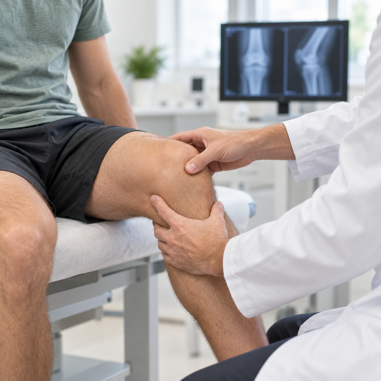
The diagnosis begins with a discussion about the circumstances of the injury, the location of the pain, and the progression of symptoms. The doctor then examines the mobility and stability of the knee. Specific tests are used to assess the medial collateral ligament and compare its stability with that of the other knee.
The assessment also looks for possible damage to the anterior cruciate ligament, the posterior cruciate ligament, a meniscus or a bone structure.
X-ray
X-rays may be performed to look for a fracture or associated bone damage.
MRI
MRI allows examination of the medial collateral ligament, assessment of the severity of the sprain, and detection of other ligament or meniscal injuries.
Ultrasound
In certain situations, ultrasound can complement the examination of the soft tissues around the knee.
The choice of examination depends on the trauma and the results of the clinical examination.
Find more information on knee imaging exams and on indications for knee MRI.
RELIEVING KNEE PAIN WITHOUT SURGERY
Most isolated sprains of the medial collateral ligament are treated without surgery. The goal is first to protect the knee, then to gradually restore its mobility, strength, and stability.
The treatment may involve several steps.
Temporarily adapt activities
Painful movements, changes of direction, and activities that put significant stress on the knee may be temporarily limited.
Protect the ligament
An articulated orthosis may be offered when the doctor believes it is useful to protect the ligament while allowing progressive knee mobility.
Gradually resume support
The resumption of weight-bearing and walking depends on the pain, stability, and severity of the sprain.
Rehabilitating the knee
Physiotherapy allows for the gradual recovery of mobility, muscle strength, balance and control of the knee.
The pace of recovery varies for each patient. The program is tailored to the clinical examination, the evolution of symptoms, and the activities the person wishes to resume.

WHEN TO CONSIDER SURGERY?
Surgery is rarely necessary for an isolated sprain of the medial collateral ligament. However, it may be considered in certain specific situations:
- very severe sprain; ;
- persistent instability despite treatment; ;
- injury that does not heal properly; ;
- damage to several ligaments; ;
- associated injury to the meniscus or another structure of the knee.
The decision depends on the clinical examination, imaging results, knee stability, and the patient's goals.
The importance of the sprain
The ligament can be stretched, partially torn, or completely ruptured.
Knee stability
Persistent instability may alter the treatment strategy.
The patient's activities and goals
The treatment takes into account the level of activity, professional constraints and the activities that the patient wishes to resume.
Resuming activities after a LCM sprain
The resumption of activities is gradual, depending on the evolution of pain, mobility, strength and stability of the knee.
Daily activities are generally resumed before more demanding movements. Walking, stairs, and physical activities are reintroduced gradually.
Resuming activities involving pivoting, changes of direction, or contact requires a specific assessment. The knee must have regained sufficient stability and good control.
Persistent pain, a feeling of instability or discomfort when resuming activity should lead to a reassessment of the knee before increasing the intensity of activities.


A CARE APPROACH TAILORED TO EACH PATIENT
Each sprain of the medial collateral ligament is different. Treatment depends on the severity of the injury, the stability of the knee, any associated injuries, and the activities the patient wishes to resume.
The assessment helps determine whether conservative treatment is sufficient or if further examinations are necessary.
The follow-up gradually accompanies the recovery and allows the different stages to be adapted: ligament protection, regaining mobility, muscle strengthening and return to activities.
For a recent knee injury, also consult the information on SOS Knee Geneva.