
FEMORACETABULAR BLUNDER
THE femoroacetabular impingement This corresponds to an abnormal or premature contact between the femur and the pelvic rim during certain movements. It may be associated with hip pain, but a bone abnormality visible on an X-ray does not necessarily cause symptoms.
Treatment is considered when pain, clinical examination, and imaging findings are consistent. Non-surgical measures are generally investigated first. Surgery may be discussed in certain situations, following an individualized assessment.
UNDERSTANDING FEMOROACETABULAR IMPACTS
 cam morphology pinching morphology
-
Normally, the head of the femur is perfectly round and extends into a thinner neck. Here, the junction between the head and the neck has a bony prominence. During significant flexion or rotation, this prominence impinges against the rim of the acetabulum.
-
Normally, the acetabulum covers the femoral head just enough. Here, its rim is too prominent or too covering. In certain positions, it touches the femoral neck earlier than intended and pinches the joint.
-
The acetabulum is the socket in the pelvis that receives the femoral head. Normally, its depth and orientation allow the hip to move freely. When it is too deep or incorrectly oriented, it covers the head too much: this is the origin of the "pinching" morphology.
-
This fibrocartilaginous ring borders the acetabulum and seals the hip joint. When the bones repeatedly rub against each other, it can become trapped and may become irritated or crack. However, damage to this ring can have other possible causes.
-
Repeated contact gradually wears down the cartilage, which normally ensures the smooth gliding of the joint. Over time, this wear can contribute to osteoarthritis. However, a cambered or pinched joint does not always cause pain or injury.
RECOGNIZING THE SYMPTOMS
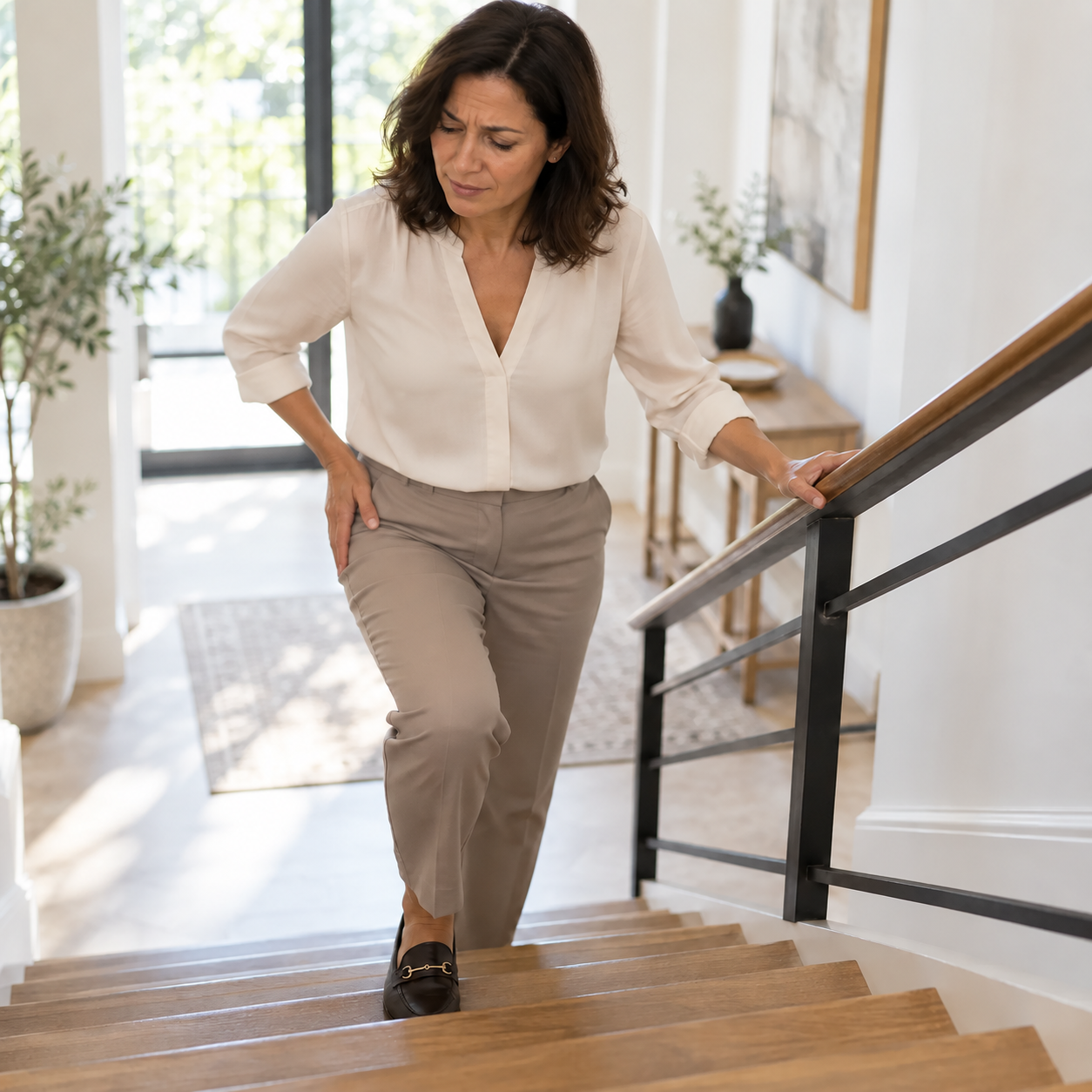
THE Symptomatic femoroacetabular impingement It most often causes deep pain in the groin or the front of the hip. It can occur during repetitive movements or positions that cause the hip to flex and rotate.
- Groin pain: during a deep bend, a squat, climbing stairs or a sporting activity.
- Discomfort after prolonged positioning: especially after sitting for a long time, depending on the person.
- Limited or uncomfortable movement: difficulty bringing the knee towards the torso or rotating the hip.
- Sensation of clicking or snapping: sometimes present, but not sufficient to confirm a labral injury or impingement.
Symptoms vary from person to person. Groin pain can also originate from the labrum, a tendon, the back, or another structure in the hip: examination helps to determine the cause.
WHY DOES FEMOROACETABULAR CONFLICT OCCUR?
The problem is related to the shape of the bones that make up the joint and how they come together during certain movements. This condition can develop during growth, particularly during periods of intense physical activity, but its origin is not always identifiable. Many people with a cambered or pinched joint experience no pain.
CAM MORPHOLOGY
A bony bump at the junction of the femoral head and neck can reduce the available space during significant flexion or rotation. It can contribute to symptoms if the examination and imaging are consistent.
MORPHOLOGY PINCH
A more prominent acetabular rim can come into contact with the femur in certain positions. This configuration can exist alone or with a cambered morphology.
MIXED FORMAT
Both morphologies are sometimes present together. Their radiological significance alone is insufficient to determine the discomfort experienced or the necessary treatment.
LABRUM: SEVERAL POSSIBLE CAUSES
The labrum can be injured in the presence of impingement, but also in other situations, such as dysplasia, trauma, instability, or joint wear. Therefore, an abnormal image alone is not sufficient to automatically link a lesion to impingement.
MAKE THE RIGHT DIAGNOSIS
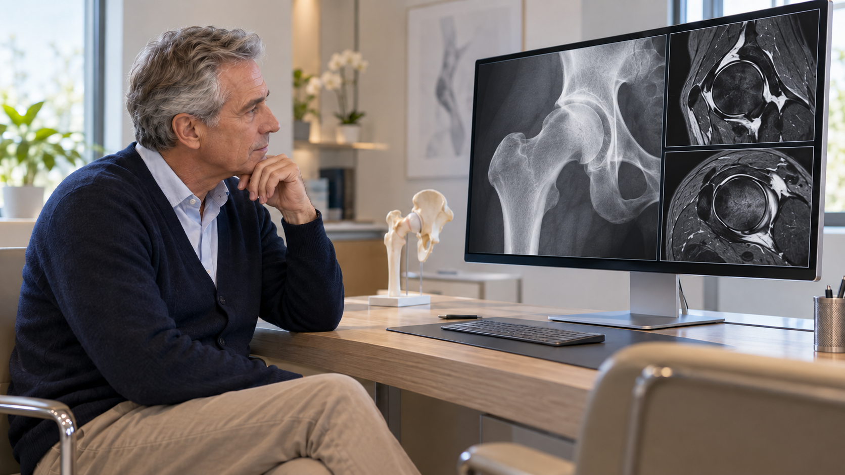
The diagnosis of femoroacetabular impingement syndrome diagnosis is not based solely on an X-ray. It combines compatible symptoms, signs observed during examination, and images showing a corresponding morphology.
01 – THE CLINICAL EXAMINATION
The doctor specifies the location and circumstances of the pain, the activities involved, and the patient's medical history. They assess gait, mobility, and movements that reproduce the symptoms. No single test is sufficient to make a diagnosis.
02 – X-RAYS
Targeted X-rays allow observation of the shape of the femur and acetabulum, as well as the overall condition of the joint. They help to identify cambered, pinched, or mixed morphology and to check for the absence of any other visible abnormalities.
03 – MRI OR CT SCAN IF NECESSARY
An MRI can examine the labrum, cartilage, and tissues surrounding the joint. A CT scan can clarify the bone structure in certain situations, particularly when preparing for surgery. These additional examinations are chosen based on the clinical question.
A morphology visible on imaging does not by itself constitute a syndrome: symptoms and examination must also be taken into account.
Relieve hip pain without surgery
When symptoms are consistent with impingement, conservative management is generally the first approach. This aims to reduce painful movements, maintain function, and gradually improve hip control.
PHYSIOTHERAPY AND STRENGTHENING
Physiotherapy can work on tolerated mobility, hip and trunk muscle strength, and movement control. The program is tailored to the individual's symptoms and goals; it does not aim to force the hip into a painful range of motion.
→ Discover the importance of physiotherapy in hip management
TEMPORARY ADAPTATION OF ACTIVITIES
It can be helpful to temporarily reduce positions that clearly trigger pain, such as repetitive deep bending or certain sports movements. The goal is to maintain a tolerated level of activity, then gradually reintroduce it as the condition improves.
DRUG TREATMENT
A pain reliever or anti-inflammatory drug may sometimes be considered for a limited period, taking into account medical history, contraindications, and other treatments. The choice and duration should be determined in consultation with a healthcare professional.
INFILTRATION IN CERTAIN SITUATIONS
An intra-articular injection may be considered in selected situations. It can help to pinpoint the articular origin of the pain or to provide temporary relief, but it does not alter the shape of the bones or repair the labrum.
The first step is to adapt the care to the symptoms and to preserve activities that remain comfortable.

WHEN TO CONSIDER SURGERY?
Surgery may be considered if the pain remains bothersome despite appropriate conservative management, if the examination and imaging are consistent, and if the condition of the joint suggests a potential benefit. The presence of a camber or pinched joint without symptoms is not an indication for surgery.
CORRECTING MORPHOLOGY
CAM
When indicated, arthroscopy can be used to smooth out the bony bump at the junction between the femoral head and neck. The procedure is planned to correct the abnormal contact while preserving as much of the bone and healthy tissue as possible.
CORRECT THE MORPHOLOGY PINCH
In some cases, the acetabular rim can be reshaped in a targeted manner. The procedure depends on the shape of the acetabulum and the coverage of the femoral head; excessive correction could compromise joint stability.
TREAT THE LABRUM IF NECESSARY
Depending on the condition and quality of the labrum, repair may be considered during the procedure. Treatment of the labrum is determined based on the findings and the context: an associated injury does not necessarily mean that it was caused by the impingement.
KEEP MOVING AND RECOVERING
In the presence of symptoms, it is not always necessary to stop all activity. Well-tolerated movements can be maintained by temporarily adjusting the range, duration, or intensity.
Physiotherapy helps individuals gradually regain the movements that are important to them by working on mobility, strength, and movement control. Progress depends on the hip's response and the established goals.
After arthroscopy, weight-bearing, crutches, and exercises are adapted to the movements performed. Recovery takes place in stages; the return to running, pivoting, or demanding sports is gradual, depending on the progress and instructions of the healthcare team.
The resumption of activities depends on the symptoms, the actions taken, and the recovery of each person.


A CARE APPROACH TAILORED TO EACH PATIENT
Two people can present with similar images and experience very different symptoms. Some bone structures never cause problems, while hip pain can have several causes. The assessment takes into account each individual's activities, clinical examination, images, and expectations.
Dr. Benoît Coulin favors a step-by-step approach: identifying the cause of the pain, starting with the least invasive and most appropriate measures, and reassessing their effectiveness. If symptoms persist and the criteria are met, intervention can be discussed with the patient.
«"An image alone is not enough to explain pain: it must be considered in relation to the symptoms, the examination, and the person's plans."»
FREQUENTLY ASKED QUESTIONS
DO YOU WANT TO BETTER UNDERSTAND YOUR PAIN?
A clinical evaluation and appropriate examinations will help to identify the source of the pain and discuss treatment options for your hip.
