

SEQUELAE AFTER A FRACTURE AND DELAYED BODY RECONSTRUCTION
After a fracture, bone healing can take longer than expected. Pain, stiffness, or functional limitations may also persist once the bone has healed: these are known as sequelae.
Treatment aims to identify the cause of the discomfort and support recovery. It depends on the condition of the bone, the symptoms, and their impact on activities.
UNDERSTANDING DELAYED BODY HEALING AND THE AFTERMARIES AFTER A FRACTURE

-
This is the area located on the inner side of the knee and is the most frequently affected by osteoarthritis. Its wear and tear can cause internal pain and gradually lead to leg deformity.
-
Located on the outer side of the knee, this compartment is less frequently affected. Its wear and tear can cause lateral pain and gradually alter the leg's alignment.
-
This joint is located between the kneecap and the femur. When it is affected, pain often appears when climbing stairs, squatting, or after sitting for a long time.

RECOGNIZING THE SYMPTOMS
Symptoms may persist after a fracture or appear when healing is delayed. Their nature depends on the bone affected, the type of fracture, and the tissues involved.
Persistent pain
It can be present at rest or appear during weight-bearing and movement. Its evolution over time is important to specify.Sensitivity at the site of the old fracture
The area may remain sensitive to pressure or certain movements.Stiffness or decreased mobility
A nearby joint may be less flexible after immobilization or the protective period.Difficulty resuming activities
Walking, weight-bearing, or certain daily activities may remain limited.
These symptoms do not always mean that the bone has not healed. Other causes can explain persistent discomfort: a medical examination is necessary to determine the cause.
WHY MAY CONSOLIDATION BE DELAYED?
Healing depends on several factors related to the fracture, the bone, and the patient's overall health. Prolonged discomfort can also result from lasting effects on surrounding tissues or mobility.
A complex fracture
Some fractures are more difficult to heal due to their location, extent, or the separation of the fragments.
Insufficient stability
Excessive movement at the fracture site can disrupt callus formation. Protection and support should follow medical recommendations.
Health-related factors
Certain diseases, smoking, or nutritional factors can influence bone repair. Their role is assessed on a case-by-case basis.
After-effects following consolidation
Even after consolidation, stiffness, persistent pain, or damage to surrounding tissues may limit activities.
MAKE THE RIGHT DIAGNOSIS
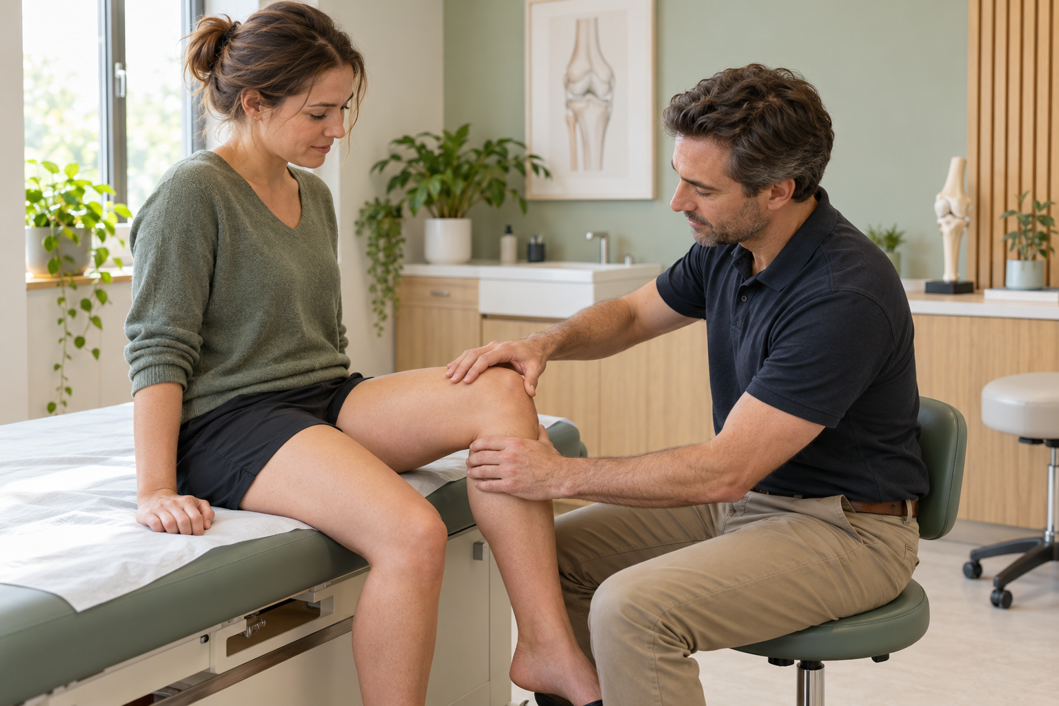
Diagnosis is based on the evolution of symptoms, clinical examination, and appropriate imaging. A single image is not sufficient to explain all persistent pain.
01 — Revisiting the history of the fracture
The doctor specifies the type of fracture, the treatments received, the weight-bearing instructions, and the evolution of the pain since the accident.
02 — Evaluate the area and the function
The clinical examination looks for localized sensitivity, limitation of movement, and difficulties encountered during functional gestures.
03 — Check the consolidation
X-rays are usually used to monitor healing. A CT scan or other imaging may be requested if the X-rays do not answer the clinical question.
The persistence of pain does not, in itself, allow us to conclude that there is a failure of consolidation.
Promoting consolidation and recovery without surgery
When the fracture remains stable and its healing process allows, medical monitoring and appropriate protection may be sufficient. The level of weight-bearing and the duration of protection are determined according to the fracture and the follow-up examinations performed.
The treatment aims to promote consolidation, control symptoms, and limit loss of mobility. It is adjusted according to clinical and radiological progress.
Adapting support and activities
Weight-bearing may be permitted, limited, or temporarily avoided depending on the location and stability of the fracture. Crutches or other walking aids may be helpful if recommended by a doctor.
Protect the fractured area
A splint, brace, or immobilizer may be maintained or adjusted as needed to protect the fracture site. These instructions should not be changed without medical advice.
Monitor the evolution
Consultations and imaging examinations allow doctors to monitor the progress of bone healing. Follow-up can also help distinguish delayed healing from pain related to surrounding tissues.
Gradually resume mobility
When permitted, physiotherapy can help restore mobility, strength, and function. Exercises are tailored to the consolidation stage and weight-bearing guidelines.
Protection and resumption of movement must be adjusted to the actual evolution of the fracture.

WHEN DOES INTERVENTION BECOME NECESSARY?
Surgery may be considered if the fracture does not progress toward sufficient healing, if it remains unstable, or if a deformity compromises function. The decision is based on imaging, physical examination, and the evolution of symptoms.
The choice also depends on the bone involved, the general state of health, treatments already carried out and the functional objectives of the patient.
Lack of progress in consolidation
If successive examinations show that consolidation is not progressing and the discomfort persists, a specialist opinion allows for discussion of possible options.
Persistent instability or movement
When the fragments remain displaced or the fracture lacks stability, intervention may be considered to restore conditions favorable to healing.
Significant functional sequelae
A persistent deformity or discomfort that severely limits weight-bearing capacity or activities may warrant surgical evaluation. All interventions are discussed on a case-by-case basis.
GRADUALLY RESUME ACTIVITIES
During the healing process, movement must adhere to protective and weight-bearing guidelines. Do not put any strain on the fractured area without medical authorization.
Recovery can begin with permitted movements and exercises, then progress to daily activities. Physiotherapy helps to work on mobility and strength when the condition of the fracture allows.
Mild discomfort may sometimes accompany the resumption of activity. A significant or persistent increase in pain, swelling, or new difficulty bearing weight should prompt adjustments to the activity and a medical consultation.
The progression takes into account follow-up examinations, symptoms, and functional recovery. More demanding activities are resumed according to the doctor's advice.
The recovery is built in stages, respecting consolidation and medical instructions.


A CARE APPROACH TAILORED TO EACH PATIENT
Recovery after a fracture varies from person to person. Persistent pain can be related to bone healing, but also to mobility or surrounding tissues.
Dr. Benoît Coulin takes into account the symptoms, function, age of the fracture, the patient's activities, and the results of the clinical examination and imaging. These elements help to determine the origin of the discomfort.
Treatment is discussed based on the progression of the fracture and the recovery goals. Monitoring and conservative measures are preferred when appropriate; intervention may be considered in certain situations.
«"The goal is to understand what limits recovery and to choose a treatment plan tailored to each fracture."»
FREQUENTLY ASKED QUESTIONS
Is your fracture still painful or hindering your activities?
A clinical examination and, if necessary, appropriate imaging can help to clarify the evolution of the fracture and the origin of the symptoms, and then to discuss a personalized treatment plan.
