

Acetabular fracture
L'acetabulum, The acetabulum, also called the acetabulum, is the socket in the pelvis into which the head of the femur fits. It directly contributes to hip stability and the transmission of loads during walking. A fracture can affect the rim of the acetabulum, one of its columns, or the upper part that bears the body's weight.
In young adults, acetabular fractures often occur after significant trauma, such as a road traffic accident or a fall from a height. In older individuals, a less severe trauma may be sufficient when the bone is weakened. The fracture may occur in isolation or in conjunction with a hip dislocation and other injuries to the pelvis or limbs.
Initial management typically begins as an emergency. Its aim is to control pain, identify any associated injuries, verify the position of the femoral head, and precisely analyze the fracture using imaging. Depending on the displacement of the fragments, hip stability, involvement of the weight-bearing surface, and the patient's overall condition, treatment may be non-surgical or require fixation.
UNDERSTANDING ACETABULUM FRACTURE

RECOGNIZING THE SYMPTOMS
A fracture of the acetabulum It usually causes significant pain in the hip, groin, or pelvis after an injury. Movement is limited, and weight-bearing may become impossible, especially when the fracture is displaced or associated with a dislocation.
- Severe hip or groin pain: It can extend towards the pelvis, buttock or thigh and be aggravated by the slightest movement.
- Difficulty or impossibility of gaining support: Walking is often very painful, or even impossible, after the trauma.
- Reduced hip mobility: Leg flexion, rotation, or extension movements may be severely limited.
- Deformity or abnormal position of the limb: An apparent rotation of the leg or shortening may suggest an associated dislocation, but the absence of visible deformity does not rule out a fracture.
After a significant injury, pelvic pain or the inability to walk should be assessed promptly. Do not force weight-bearing or attempt to mobilize or reposition the hip yourself.
WHY DOES AN ACETABULUM FRACTURE OCCUR?
An acetabular fracture occurs when the forces exerted on the pelvis exceed the bone's strength. The mechanism and bone density influence the shape of the fracture, its displacement, and the treatment strategy.
HIGH-ENERGY TRAUMA
A road accident, a fall from a height, or a violent sports impact can transmit significant force to the hip. The resulting injuries can be complex and associated with other fractures or trauma.
FALL IN AN ELDERLY PERSON
With age, a simple fall can cause a fracture of the acetabulum when the bone is weakened. The assessment must then take into account osteoporosis, the individual's level of independence, and other health problems.
DIRECT IMPACT OR TRANSMITTED VIA THE FEMUR
A lateral impact to the hip or a force transmitted by the flexed femur can fracture the rim or columns of the acetabulum. Hip dislocation may be associated.
BONE FRAGILITY OR PRE-EXISTING INJURY
Osteoporosis, bone disease, or, more rarely, an injury weakening the pelvis can predispose to fracture. A fracture resulting from moderate trauma may prompt an investigation for bone fragility.

MAKE THE RIGHT DIAGNOSIS
Diagnosis is based on clinical examination and imaging. Pelvic X-rays provide an initial view of the fracture, but a CT scan is very often necessary to analyze the columns, fragment displacement, weight-bearing surface, and hip congruity.
01 – THE CLINICAL EXAMINATION
The doctor will look for pain, limited range of motion, any deformities, and weight-bearing capacity. They will also check sensation, motor function, and circulation in the lower limb, as well as any signs suggesting an associated injury.
02 – X-RAYS
X-rays of the pelvis and hip can be used to look for fractures, dislocations, and significant displacement. Specific views can be taken to better analyze the contours of the acetabulum.
03 – THE SCANNER AND THE OVERALL ASSESSMENT
The scanner provides cross-sectional analysis and 3D reconstructions of the fracture. It helps in treatment planning and in assessing the condition of the femoral head. Depending on the situation, preoperative, laboratory, and anesthetic assessments are also performed.
An inconclusive X-ray does not rule out an acetabular fracture: if pain, limited movement, or inability to bear weight persists, a CT scan may be necessary.
TREATING THE FRACTURE AND PRESERVING THE HIP
The management of a fracture of the acetabulum The outcome depends on hip stability, fragment displacement, involvement of the weight-bearing area, and the patient's overall condition. The goal is to restore a stable and as regular a joint as possible, while minimizing complications related to trauma and immobilization.
RELIEVE PAIN AND PROTECT THE HIP
The limb is placed in a comfortable position, and weight-bearing is avoided until the fracture has been diagnosed. Pain medication is administered, and the patient's general condition, circulation, and sensation are monitored. In the event of a dislocation, urgent reduction may be required under medical supervision.
TREAT WITHOUT SURGERY WHEN THE FRACTURE IS STABLE
A minimally displaced or non-displaced fracture, with a stable hip and sufficiently preserved joint surface, can sometimes be treated without surgery. Management involves limited or protected weight-bearing, walking aids, radiographic monitoring, and a gradual return to activity depending on the patient's progress.
REDUCE AND FIX THE FRACTURE IF NECESSARY
When bone fragments are displaced, the weight-bearing surface is irregular, or the hip is unstable, surgery may be recommended. Depending on the fracture, the surgeon will realign the fragments and fix them with screws, plates, or percutaneous fixation when appropriate. The choice depends on the fracture's morphology and the bone quality.
MONITORING CONSOLIDATION AND HIP CONDITION
Follow-up care checks for bone healing, fragment positioning, hip stability, and weight-bearing. Monitoring is also necessary to detect stiffness, femoral head necrosis, post-traumatic osteoarthritis, phlebitis, or infection after surgery.
The treatment is not just about strengthening the bone: it aims to preserve a stable, mobile and functional hip in the long term. The strategy is tailored to the fracture, the age, the autonomy and the expectations of the patient.
IS SURGERY NECESSARY?
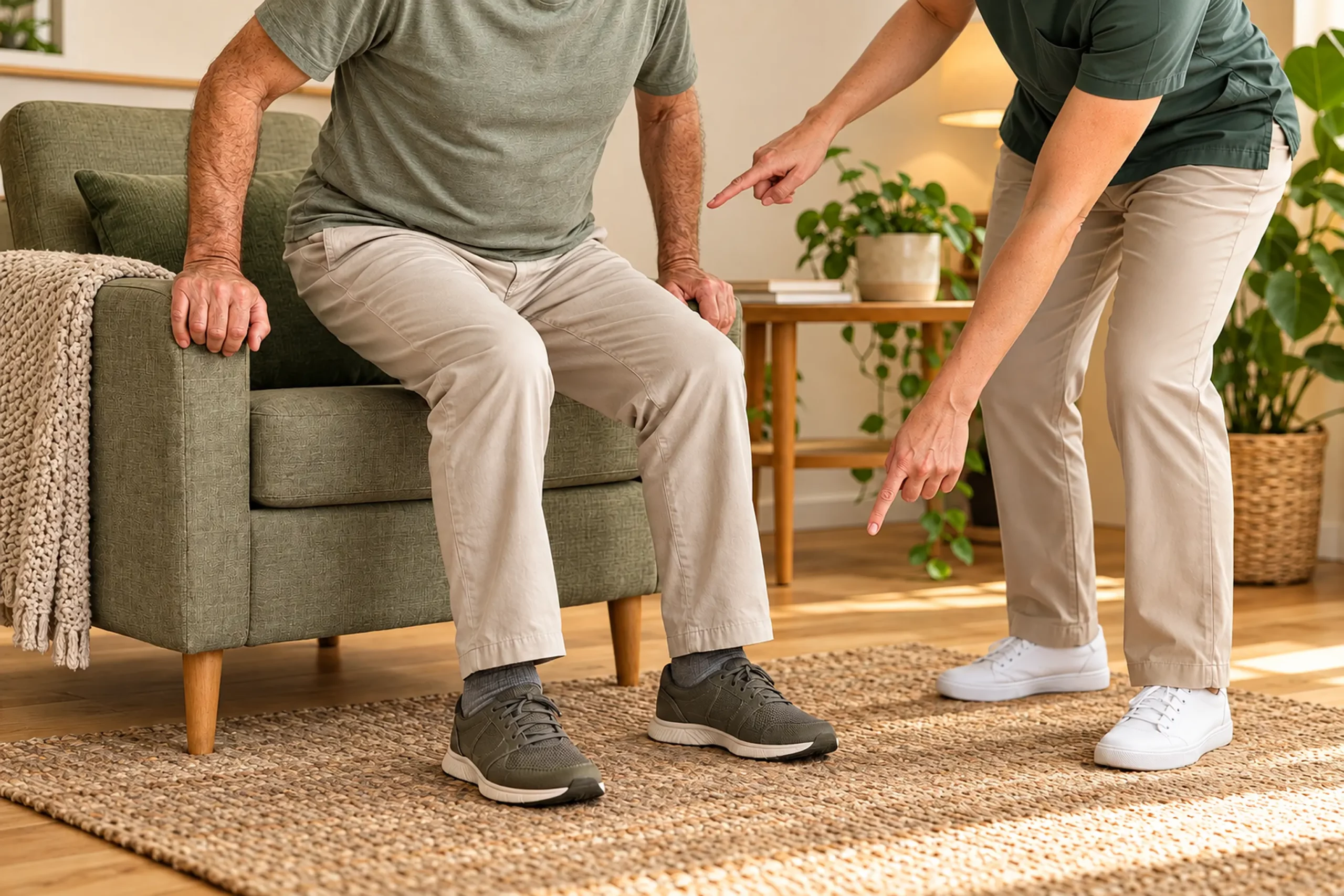
Surgery is not always necessary for all acetabular fractures. It is primarily considered when the fracture is displaced, involves the weight-bearing area, the hip is unstable, or the femoral head is no longer properly centered. A stable, minimally displaced fracture can sometimes be monitored and treated with protected weight-bearing.
RESTORE HIP CONGRUENCE
When displaced fragments make the surface of the acetabulum irregular, reduction and fixation may be necessary to restore the best possible contact between the femoral head and the acetabulum.
STABILIZING A DISPLACED FRACTURE OR ASSOCIATED DISLOCATION
An unstable hip, a dislocation or a large fragment of the acetabular rim may warrant intervention to secure the joint and limit abnormal stresses.
TAKE INTO ACCOUNT THE GENERAL CONDITION AND BONE QUALITY
Age, associated medical conditions, pre-injury mobility, bone quality, and anesthetic risks are all factors considered in the decision. In some patients, an alternative strategy may be discussed if conventional fixation presents too high a risk or if the joint is already severely damaged.
REHABILITATION AND RESUMPTION OF ACTIVITIES
Recovery depends on the type of fracture, hip stability, the chosen treatment, and the patient's overall health. Rehabilitation begins as soon as the situation is sufficiently stable. It can continue in the hospital, at a rehabilitation center, or at home.
01 – CONTROL PAIN AND MOBILIZE SAFELY
The initial repositioning is carried out with the care team. Transfers and movements take into account pain, balance, and permitted weight-bearing.
02 – RESUME SUPPORT WITH AID
Walking begins with crutches, a frame, or a walker when permitted. The level of weight-bearing depends on the fracture, its healing, and the stability of the fixation.
03 – RECOVER MOBILITY AND STRENGTH
Physiotherapy gradually works on hip mobility, pelvic and thigh muscles, transfers and balance, without exceeding protective guidelines.
04 — MONITOR THE CONSOLIDATION
Clinical and radiographic checks allow for adjustments to support and exercises. Progress does not follow the same schedule for everyone.
05 — RESUME DAILY ACTIVITIES
Personal hygiene, dressing, using stairs, and mobility are relearned or adapted. Resumption of sports and demanding activities is discussed after sufficient recovery and consolidation of injuries.
The resumption of weight-bearing and activities depends on the consolidation and stability of the hip, and not on an identical timeframe for all patients.

A CARE APPROACH TAILORED TO EACH PATIENT
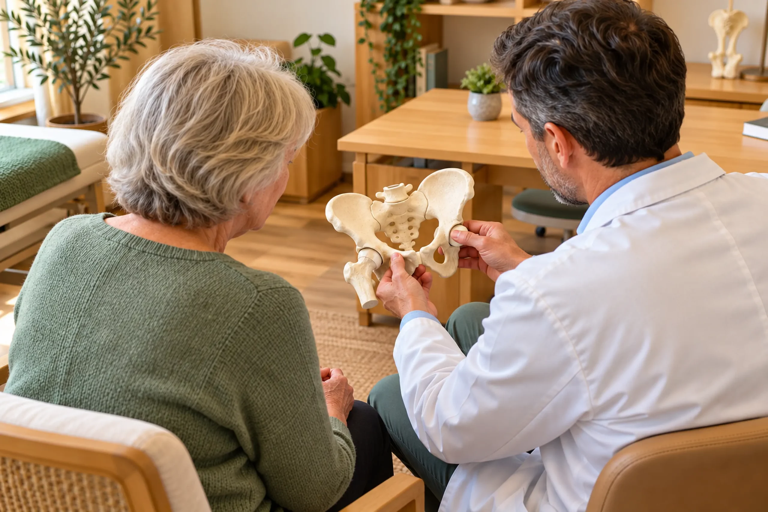
An acetabular fracture cannot be treated solely based on an X-ray. The decision takes into account the location of the fracture, the displacement of the fragments, the involvement of the weight-bearing area, the stability of the hip, the condition of the cartilage, and the quality of the bone.
Management involves pain control, weight-bearing protection, assessment for associated injuries, and monitoring of bone healing. When surgery is indicated, the strategy is chosen based on the fracture anatomy and the patient's overall condition.
After the acute phase, follow-up checks mobility, stability, healing, and the gradual return to activities. Prolonged monitoring can be useful to look for post-traumatic osteoarthritis or other hip complications.
«"Treatment should preserve hip congruity as much as possible and gradually support functional recovery."»
FREQUENTLY ASKED QUESTIONS
DO YOU WANT TO BETTER UNDERSTAND YOUR PAIN?
A clinical examination combined with appropriate imaging helps to determine the origin of symptoms and identify the most suitable treatments.
