

Patellar instability or dislocation
There patellar dislocation This occurs when the patella slips out of its normal position, usually towards the outside of the knee. It may return to its normal position on its own or require medical reduction. Pain and swelling are common after the episode.
A first dislocation does not automatically lead to surgery. Treatment depends in particular on associated injuries, the shape of the joint, the stability of the knee and the risk of recurrence.
UNDERSTANDING BALL JOINT MOVEMENT

-
This is the area located on the inner side of the knee and is the most frequently affected by osteoarthritis. Its wear and tear can cause internal pain and gradually lead to leg deformity.
-
Located on the outer side of the knee, this compartment is less frequently affected. Its wear and tear can cause lateral pain and gradually alter the leg's alignment.
-
This joint is located between the kneecap and the femur. When it is affected, pain often appears when climbing stairs, squatting, or after sitting for a long time.

RECOGNIZING THE SYMPTOMS
A dislocation may be obvious when the kneecap remains displaced, but it may also have returned to its original position before the examination. Pain, swelling, and a feeling of instability may then persist.
- Pain in the front of the knee: It often appears suddenly at the time of movement and can increase when the knee is moved or put under load.
- Swelling : The knee can swell rapidly after the episode, sometimes significantly.
- Visible deformation: If the kneecap is still dislocated, it may appear to be displaced to the side of the knee.
- Difficulty bending or straightening the leg: Pain and swelling can limit movement.
- The feeling that the knee is going to give way: After a dislocation, some people worry that the kneecap may dislocate again, especially when pivoting or bending the knee.
After an initial episode, a feeling of apprehension or instability may persist. Repeated episodes suggest patellar instability, for which the anatomical factors and any associated injuries should be investigated.
A kneecap that is still displaced, severe pain, marked swelling, or the inability to bear weight warrants prompt evaluation. A persistent deformity should not be attempted by the patient themselves. In case of an acute injury, consult the emergency department.’knee emergencies.
WHY CAN THE PALMULE DISLOCATE?
A dislocation occurs when the kneecap moves out of its normal position within the trochlea. It often happens during a pivoting motion, a change of direction, landing after a jump, or an impact. The cause may combine the traumatic movement with specific characteristics of the joint.
A twisting motion or an impact
The foot may remain fixed to the ground while the body rotates, or the knee may be subjected to an impact. The kneecap is then forced outward. This episode can cause damage to the MPFL and sometimes cartilage lesions or an osteochondral fragment.
The shape of the joint
A shallow trochlea, a relatively high-set patella, or certain lower limb alignment parameters can make patellar guidance less stable. These characteristics do not necessarily cause dislocation on their own, but they can contribute to the risk of recurrence.
Laxity or a history
Ligament laxity, a previous anterior dislocation, or pre-existing instability may also be considered in the assessment. Risk cannot be determined from a single factor; the examination and imaging are interpreted together.
Repeated episodes can maintain apprehension and sometimes damage the cartilage. The assessment may also look for a knee cartilage damage if the symptoms or imaging suggest it.
MAKE THE RIGHT DIAGNOSIS
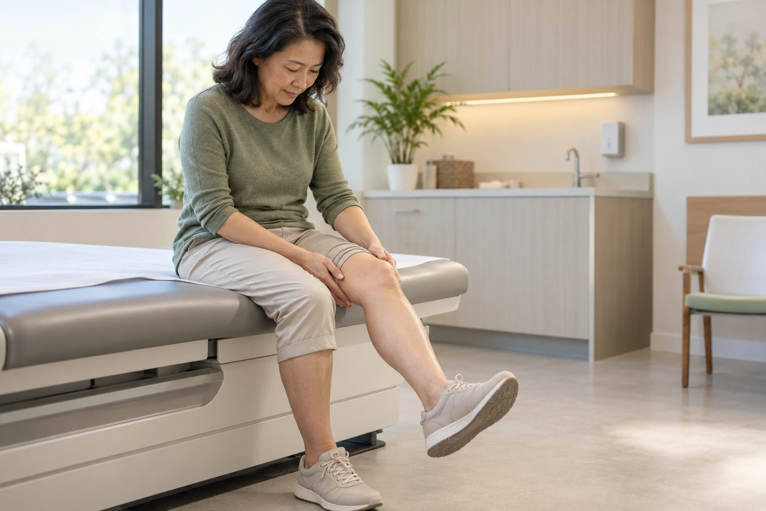
The assessment determines whether the kneecap is still displaced, whether it has returned to its normal position, and whether any other structures of the knee have been affected. It takes into account the circumstances of the episode and any history of instability.
01 — THE CIRCUMSTANCES OF THE EPISODE
The doctor asks how the knee moved, whether the kneecap spontaneously returned to its place, whether reduction was necessary, and whether a similar episode has occurred before.
02 — THE CLINICAL EXAMINATION
The examination assesses pain, swelling, mobility, and stability, taking into account the patient's tolerance. It also looks for signs that may suggest another knee injury.
03 — X-RAYS
Depending on the context, X-rays can be used to observe the position of the kneecap, its alignment, and the possible presence of a fracture or bone fragment.
04 — MRI OR ANOTHER TARGETED EXAMINATION
An MRI can help assess the MPFL, cartilage, and associated lesions, particularly after trauma with significant swelling. A CT scan can be useful in some cases to clarify bone parameters or prepare for possible surgery.
Simply putting the kneecap back in place is not enough to rule out an associated injury.
INITIAL CARE
The initial steps aim to protect the knee and assess the situation. Treatment depends on the position of the kneecap, the pain, and any suspected injuries.
Protect the knee
Avoid putting strain on a painful leg or attempting to reposition a dislocated kneecap yourself. If it remains visibly dislocated, urgent evaluation is necessary. Crutches or a protective brace may be recommended temporarily depending on the pain and stability of the leg.
To reduce pain and swelling
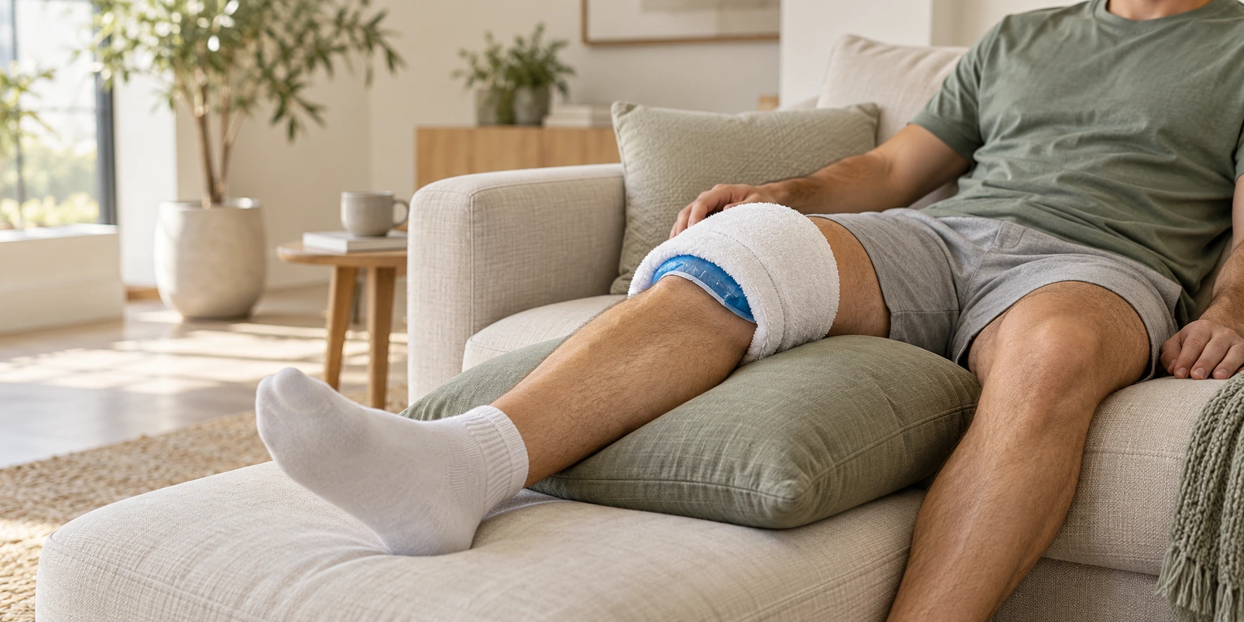
Relative rest, elevating the leg, and applying cold compresses wrapped in a cloth can help reduce discomfort. Pain medication can be discussed with a healthcare professional depending on the patient's medical condition.
Organize an exam
Pain relievers or anti-inflammatory treatments may be used during certain painful periods, depending on medical history and any contraindications.
They help control symptoms, but do not replace appropriate activity, muscle strengthening, and addressing mechanical factors.
CONSERVATIVE TREATMENT: STABILIZE AND REHABILITATE
After the first episode, non-surgical treatment is often considered when the kneecap is back in place and no associated injuries require surgery. The decision depends on the assessment and the patient's profile.
Gradually resume the movements
Temporary protection may sometimes be recommended, but its usefulness and duration depend on the examination. The goal is to gradually regain mobility without unnecessarily immobilizing the knee.
Working on strength and control
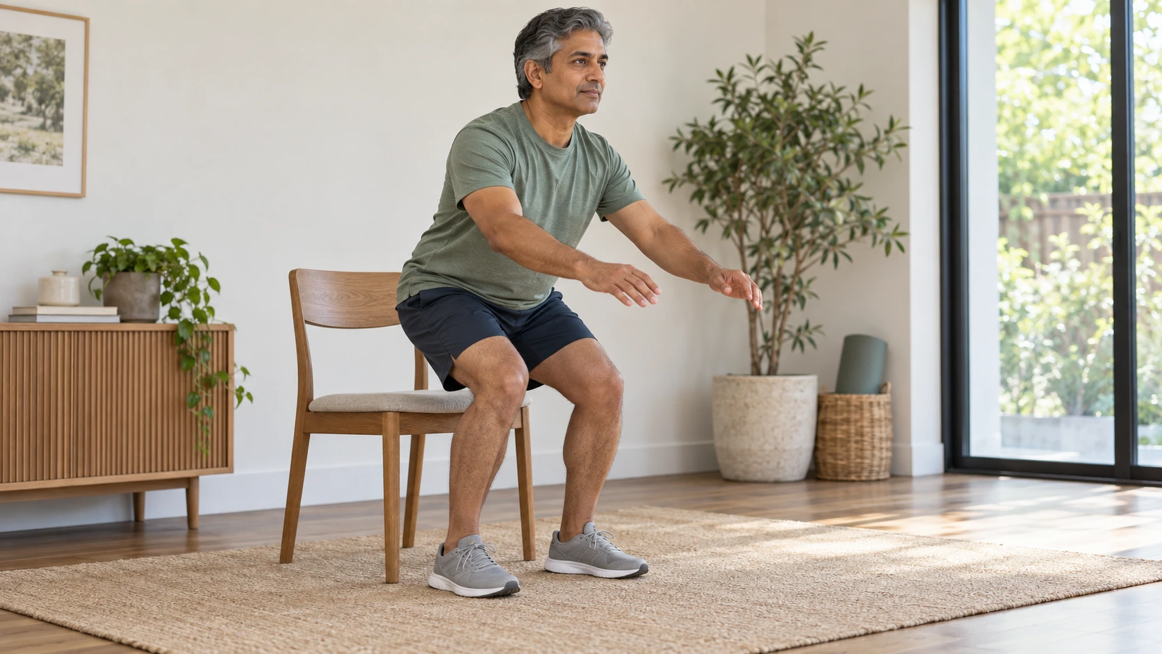
Physiotherapy helps to regain range of motion, strengthen thigh and hip muscles, and improve lower limb control. Exercises are adjusted according to pain, swelling, and stability.
Adapt activities and monitor developments
Movements that trigger pain or apprehension are temporarily modified. Walking and other activities are resumed gradually, depending on clinical progress. Re-evaluation is indicated if the knee remains unstable, if episodes recur, or if recovery stalls.
A well-monitored conservative approach may be appropriate after a first episode, but it must be tailored to the lesions and the risk of recurrence.
To better understand the role of rehabilitation, consult the page on the knee rehabilitation.
SURGICAL TREATMENT: WHEN STABILITY REMAINS THREATENED
Surgery is not always necessary after a first dislocation. It may be considered in cases of repeated dislocations, persistent instability despite appropriate rehabilitation, or osteochondral lesions requiring specific treatment. The assessment aims to identify the factors contributing to the displacement.
GESTURES ADAPTED TO ANATOMY
Depending on the case, medial patellofemoral ligament reconstruction may be recommended to improve stability. Bone correction may be considered if certain anatomical factors contribute to instability. The choice of procedure depends on the clinical assessment and imaging: there is no single operation suitable for all patients.
PRESERVE THE STRUCTURES AS MUCH AS POSSIBLE
The intervention aims to treat the identified mechanism and reduce episodes of instability. It does not guarantee the absence of pain or recurrence and requires a period of rehabilitation. The timing of the intervention depends in particular on the swelling, associated injuries, and progress after initial treatment.
The page on the patella surgery presents the techniques and their indications.

REHABILITATION AND RESUMPTION OF ACTIVITIES
Recovery depends on the severity of the episode, any associated injuries, and the chosen treatment. The steps are adapted to the knee's progress; the timeframes do not guarantee a full recovery.
01 — Gradually regain mobility
The exercises aim to restore flexion and extension without permanently increasing pain or swelling.
02 — Reactivate and strengthen the muscles
Muscle work progresses according to tolerance, particularly to improve dynamic knee support.
03 — Control the movements
Physiotherapy works on balance, coordination and control of the knee during functional movements.
04 — Resume daily activities
Walking, stairs and usual activities are gradually reintroduced, depending on regained mobility, strength and stability.
05 — Returning to sport with functional criteria
Resuming changes of direction, jumps, and pivoting sports requires sufficient strength, good movement control, and the absence of instability. After surgery, the timeline also depends on the specific movement performed and the progress made during rehabilitation.
The decision to return to sport depends on the capabilities of the knee, not solely on the time elapsed.
There knee rehabilitation accompanies this progression.
A STRATEGY TAILORED TO EACH PATIENT
Treatment depends on the history of the knee, the number of episodes, the presence of associated injuries, and the activities the patient wishes to resume. Dr. Benoît Coulin takes into account the clinical examination, imaging, perceived stability, anatomical parameters, and each patient's individual goals.
Conservative treatment avoids intervention and prioritizes gradual recovery, but it may be insufficient when instability recurs or when an associated injury warrants it. Surgery can stabilize certain situations, but it has its own limitations and requires structured rehabilitation.
The choice is built upon the assessment and evolution, explaining the expected benefits and limitations of each option.
«"Stabilizing the kneecap begins with understanding why it moves."»

FREQUENTLY ASKED QUESTIONS
DID YOU HAVE A DISLOCATED PATELLA?
A clinical examination and imaging, chosen according to your symptoms, will help assess any associated injuries and instability factors. Dr. Benoît Coulin can discuss a treatment plan tailored to your knee and your activities.
